

Morphologies, mechanical and in vitro behaviors of DLP-based 3D printed HA scaffolds with different structural configurations
- PMID: 37441027
- PMCID: PMC10333813
- DOI: 10.1039/d3ra03080f
Morphologies, mechanical and in vitro behaviors of DLP-based 3D printed HA scaffolds with different structural configurations
Abstract
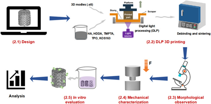
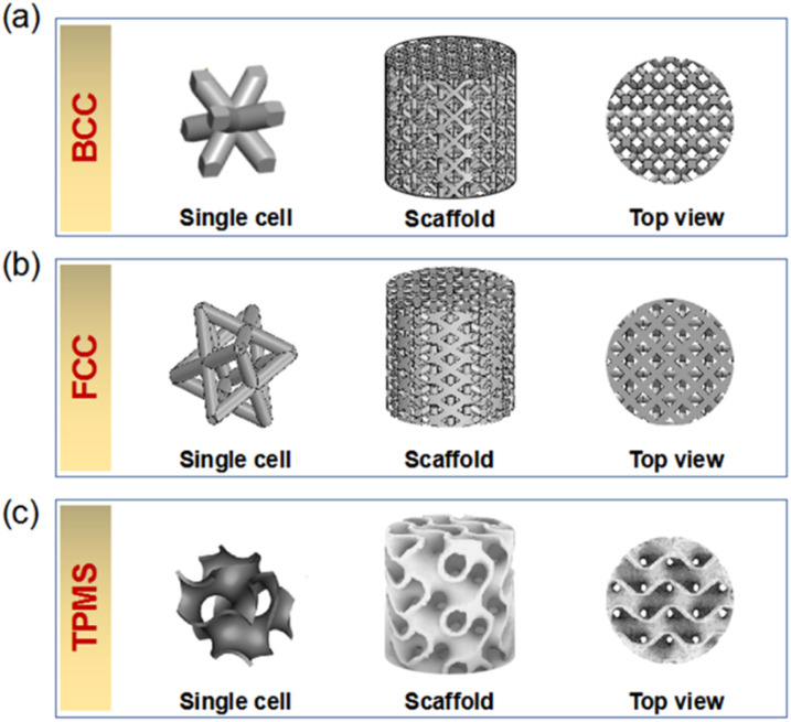
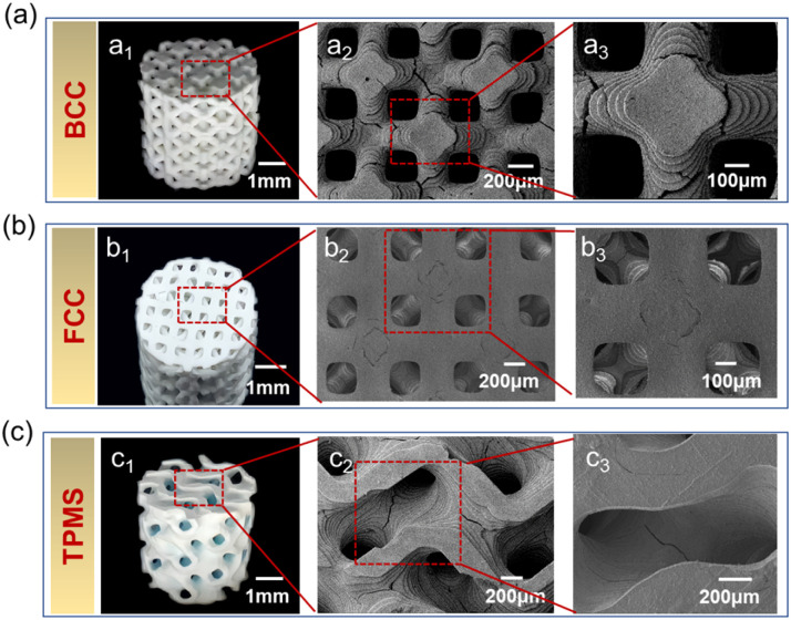
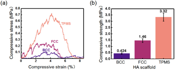
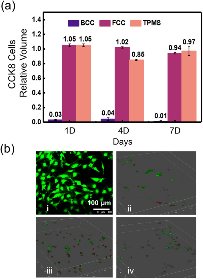
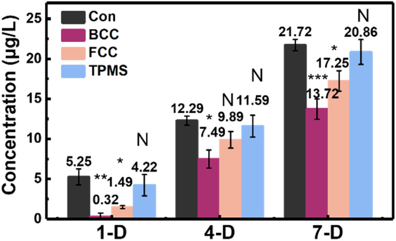
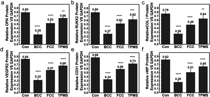
In the field of bone engineering, porous ceramic scaffolds are in great demand for repairing bone defects. In this study, hydroxyapatite (HA) ceramic scaffolds with three different structural configurations, including the body-centered cubic (BCC), the face-centered cubic (FCC), and the triply periodic minimal surface (TPMS), were fabricated through digital light processing (DLP) based 3D printing technologies. The effects of the structural configurations on the morphologies and mechanical properties of the DLP-based 3D printed HA scaffolds were characterized. Furthermore, in vitro evaluations, including in vitro cytocompatibility, bone alkaline phosphatase (ALP) activity assay, and protein expression, were conducted to assess HA scaffold behavior. Finally, we evaluated the effects of structural configurations from these aspects and selected the most suitable structure of HA scaffold for bone repair.
This journal is © The Royal Society of Chemistry.
Conflict of interest statement
There are no conflicts to declare.
Figures
Similar articles
-
Three-Dimensionally Printed Bionic Hydroxyapatite (HAp) Ceramic Scaffolds with Different Structures and Porosities: Strength, Biocompatibility, and Biomedical Application Potential.Materials (Basel). 2024 Dec 13;17(24):6092. doi: 10.3390/ma17246092. Materials (Basel). 2024. PMID: 39769691 Free PMC article.
-
Nano-Hydroxyapatite Bone Scaffolds with Different Porous Structures Processed by Digital Light Processing 3D Printing.Int J Bioprint. 2022 Jan 17;8(1):502. doi: 10.18063/ijb.v8i1.502. eCollection 2022. Int J Bioprint. 2022. PMID: 35187284 Free PMC article.
-
3D printed TPMS structural PLA/GO scaffold: Process parameter optimization, porous structure, mechanical and biological properties.J Mech Behav Biomed Mater. 2023 Jun;142:105848. doi: 10.1016/j.jmbbm.2023.105848. Epub 2023 Apr 18. J Mech Behav Biomed Mater. 2023. PMID: 37099921
-
Novel 3D printed TPMS scaffolds: microstructure, characteristics and applications in bone regeneration.J Tissue Eng. 2024 Jul 26;15:20417314241263689. doi: 10.1177/20417314241263689. eCollection 2024 Jan-Dec. J Tissue Eng. 2024. PMID: 39071895 Free PMC article. Review.
-
Three-dimensional (3D) printed scaffold and material selection for bone repair.Acta Biomater. 2019 Jan 15;84:16-33. doi: 10.1016/j.actbio.2018.11.039. Epub 2018 Nov 24. Acta Biomater. 2019. PMID: 30481607 Review.
Cited by
-
Biomaterials Adapted to Vat Photopolymerization in 3D Printing: Characteristics and Medical Applications.J Funct Biomater. 2023 Dec 22;15(1):7. doi: 10.3390/jfb15010007. J Funct Biomater. 2023. PMID: 38248674 Free PMC article. Review.
-
Fabrication Strategies for Bioceramic Scaffolds in Bone Tissue Engineering with Generative Design Applications.Biomimetics (Basel). 2024 Jul 5;9(7):409. doi: 10.3390/biomimetics9070409. Biomimetics (Basel). 2024. PMID: 39056850 Free PMC article. Review.
-
A preliminary study of cell-based bone tissue engineering into 3D-printed β-tricalcium phosphate scaffolds and polydioxanone membranes.Sci Rep. 2024 Dec 28;14(1):31184. doi: 10.1038/s41598-024-82334-6. Sci Rep. 2024. PMID: 39732806 Free PMC article.
-
How to Improve the Curing Ability during the Vat Photopolymerization 3D Printing of Non-Oxide Ceramics: A Review.Materials (Basel). 2024 May 29;17(11):2626. doi: 10.3390/ma17112626. Materials (Basel). 2024. PMID: 38893890 Free PMC article. Review.
-
Three-Dimensionally Printed Bionic Hydroxyapatite (HAp) Ceramic Scaffolds with Different Structures and Porosities: Strength, Biocompatibility, and Biomedical Application Potential.Materials (Basel). 2024 Dec 13;17(24):6092. doi: 10.3390/ma17246092. Materials (Basel). 2024. PMID: 39769691 Free PMC article.
References
-
- Bogala M. Bioprinting. 2022;28:e00244.
-
- Li Y. y. Li L. t. Li B. Mater. Des. 2015;72:16–20.
-
- Baino F. Fiume E. Barberi J. Kargozar S. Marchi J. Massera J. Verné E. Int. J. Appl. Ceram. Technol. 2019;16(5):1762–1796.
-
- Shih S.-J. Sari D. R. M. Lin Y.-C. Int. J. Appl. Ceram. Technol. 2016;13(4):787–794.
LinkOut - more resources
Full Text Sources