

Asymmetrical putaminal atrophy in parkinsonism-predominant multiple system atrophy (MSA-P): A case report
- PMID: 37441448
- PMCID: PMC10333103
- DOI: 10.1016/j.radcr.2023.05.060
Asymmetrical putaminal atrophy in parkinsonism-predominant multiple system atrophy (MSA-P): A case report
Abstract
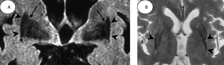
We encountered a case of multiple system atrophy parkinsonian subtype (MSA-P) with right-dominant parkinsonism in the early stage of the disease. Atrophy of the posterolateral putamen and iron deposition are the neuropathological hallmark of MSA-P. Coronal fluid-attenuated inversion-recovery (FLAIR) images showed atrophy and iron deposition in the left posterior putamen contralateral to the clinical dominant side in the early phase. Atrophy in the posterior putamen of patients with MSA-P was more clearly observed on coronal FLAIR images than on axial T2-weighted images. These findings reflected the pathological changes and might be a pathognomonic sign of MSA-P.
Keywords: Asymmetrical parkinsonism; FLAIR; Lateral medullary laminae; Putamen.
© 2023 The Authors. Published by Elsevier Inc. on behalf of University of Washington.
Figures

Similar articles
-
Differentiation of Parkinsonism-Predominant Multiple System Atrophy from Idiopathic Parkinson Disease Using 3T Susceptibility-Weighted MR Imaging, Focusing on Putaminal Change and Lesion Asymmetry.AJNR Am J Neuroradiol. 2015 Dec;36(12):2227-34. doi: 10.3174/ajnr.A4442. Epub 2015 Sep 3. AJNR Am J Neuroradiol. 2015. PMID: 26338919 Free PMC article.
-
The putaminal abnormalities on 3.0T magnetic resonance imaging: can they separate parkinsonism-predominant multiple system atrophy from Parkinson's disease?Acta Radiol. 2015 Mar;56(3):322-8. doi: 10.1177/0284185114524090. Epub 2014 Mar 11. Acta Radiol. 2015. PMID: 24619850
-
Hyperintense putaminal rim at 1.5 T: prevalence in normal subjects and distinguishing features from multiple system atrophy.BMC Neurol. 2012 Jun 18;12:39. doi: 10.1186/1471-2377-12-39. BMC Neurol. 2012. PMID: 22708511 Free PMC article.
-
Progression and prognosis in multiple system atrophy: an analysis of 230 Japanese patients.Brain. 2002 May;125(Pt 5):1070-83. doi: 10.1093/brain/awf117. Brain. 2002. PMID: 11960896 Review.
-
[Multiple system atrophy].Psychol Neuropsychiatr Vieil. 2010 Sep;8(3):179-91. doi: 10.1684/pnv.2010.0212. Psychol Neuropsychiatr Vieil. 2010. PMID: 20739256 Review. French.
Cited by
-
Two distinct degenerative types of nigrostriatal dopaminergic neuron in the early stage of parkinsonian disorders.Clin Park Relat Disord. 2024 Feb 15;10:100242. doi: 10.1016/j.prdoa.2024.100242. eCollection 2024. Clin Park Relat Disord. 2024. PMID: 38405025 Free PMC article.
-
Asymmetry in Atypical Parkinsonian Syndromes-A Review.J Clin Med. 2024 Sep 28;13(19):5798. doi: 10.3390/jcm13195798. J Clin Med. 2024. PMID: 39407856 Free PMC article. Review.
References
-
- Fanciulli A, Wenning GK. Multiple-system atrophy. N Engl J Med. 2015;372:249–263. - PubMed
-
- Koga S, Dickson DW. Recent advances in neuropathology, biomarkers and therapeutic approach of multiple system atrophy. J Neurol Neurosurg Psychiatry. 2018;89:175–184. - PubMed
-
- Osaki Y, Wenning GK, Daniel SE, Hughes A, Lees AJ, Mathias CJ, et al. Do published criteria improve clinical diagnostic accuracy in multiple system atrophy? Neurology. 2002;59:1486–1491. - PubMed
-
- Joutsa J, Gardberg M, Röyttä M, Kaasinen V. Diagnostic accuracy of parkinsonism syndromes by general neurologists. Parkinsonism Relat Disord. 2014;20:840–844. - PubMed
Publication types
LinkOut - more resources
Full Text Sources
