

Molecular insights into mineralotropic hormone inter-regulation
- PMID: 37441497
- PMCID: PMC10334211
- DOI: 10.3389/fendo.2023.1213361
Molecular insights into mineralotropic hormone inter-regulation
Abstract
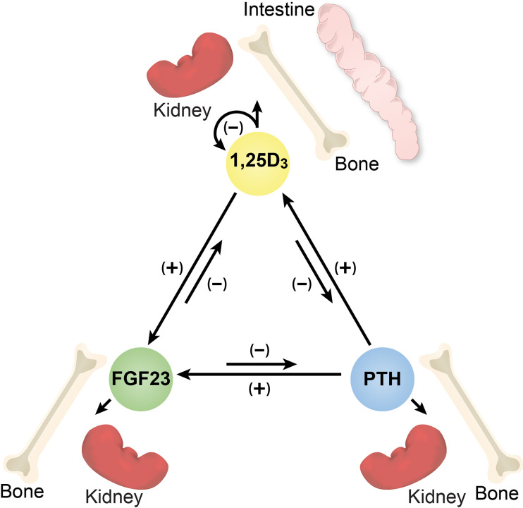
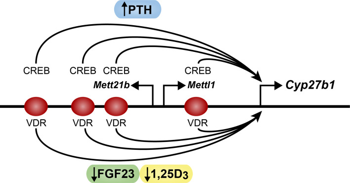
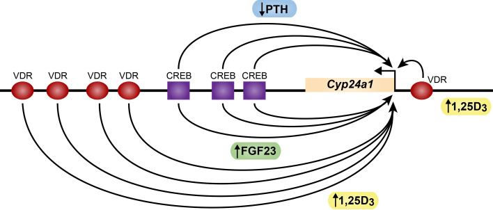
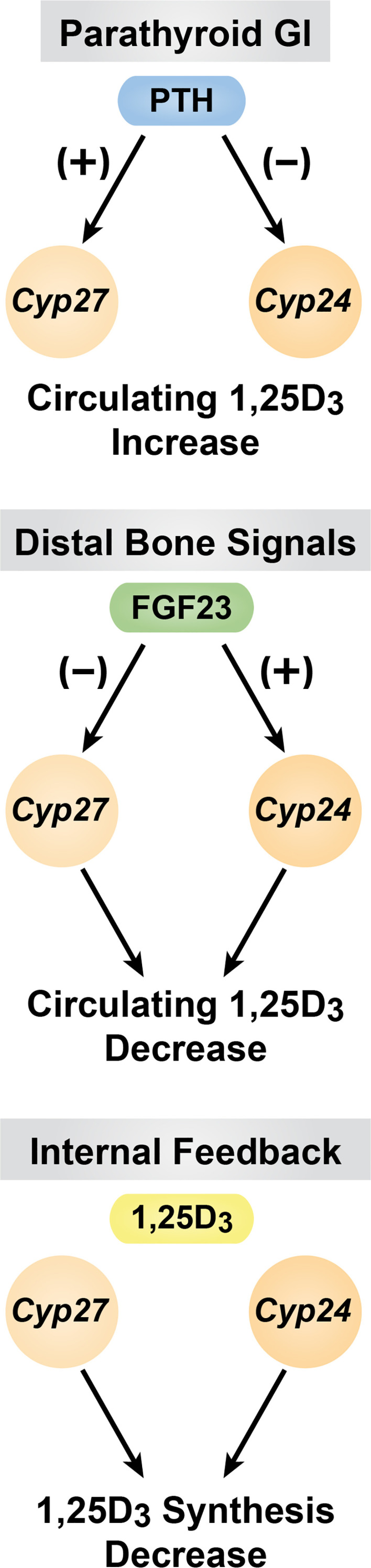
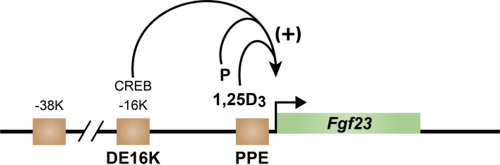
The regulation of mineral homeostasis involves the three mineralotropic hormones PTH, FGF23 and 1,25-dihydroxyvitamin D3 (1,25(OH)2D3). Early research efforts focused on PTH and 1,25(OH)2D3 and more recently on FGF23 have revealed that each of these hormones regulates the expression of the other two. Despite early suggestions of transcriptional processes, it has been only recently that research effort have begun to delineate the genomic mechanisms underpinning this regulation for 1,25(OH)2D3 and FGF23; the regulation of PTH by 1,25(OH)2D3, however, remains obscure. We review here our molecular understanding of how PTH induces Cyp27b1 expression, the gene encoding the enzyme responsible for the synthesis of 1,25(OH)2D3. FGF23 and 1,25(OH)2D3, on the other hand, function by suppressing production of 1,25(OH)2D3. PTH stimulates the PKA-induced recruitment of CREB and its coactivator CBP at CREB occupied sites within the kidney-specific regulatory regions of Cyp27b1. PKA activation also promotes the nuclear translocation of SIK bound coactivators such as CRTC2, where it similarly interacts with CREB occupied Cyp27b1 sites. The negative actions of both FGF23 and 1,25(OH)2D3 appear to suppress Cyp27b1 expression by opposing the recruitment of CREB coactivators at this gene. Reciprocal gene actions are seen at Cyp24a1, the gene encoding the enzyme that degrades 1,25(OH)2D3, thereby contributing to the overall regulation of blood levels of 1,25(OH)2D3. Relative to PTH regulation, we summarize what is known of how 1,25(OH)2D3 regulates PTH suppression. These studies suggest that it is not 1,25(OH)2D3 that controls PTH levels in healthy subjects, but rather calcium itself. Finally, we describe current progress using an in vivo approach that furthers our understanding of the regulation of Fgf23 expression by PTH and 1,25(OH)2D3 and provide the first evidence that P may act to induce Fgf23 expression via a complex transcriptional mechanism in bone. It is clear, however, that additional advances will need to be made to further our understanding of the inter-regulation of each of these hormonal genes.
Keywords: CRISPR/Cas9; ChIP-seq analysis; Cyp27b1/Cyp24a1 genes; FGF23 gene; PTH gene; mineral regulating hormones; mutant mice; transcription.
Copyright © 2023 Pike, Lee and Meyer.
Conflict of interest statement
The authors declare that the research was conducted in the absence of any commercial or financial relationships that could be construed as a potential conflict of interest.
Figures

Similar articles
-
Rapid genomic changes by mineralotropic hormones and kinase SIK inhibition drive coordinated renal Cyp27b1 and Cyp24a1 expression via CREB modules.J Biol Chem. 2022 Nov;298(11):102559. doi: 10.1016/j.jbc.2022.102559. Epub 2022 Sep 30. J Biol Chem. 2022. PMID: 36183832 Free PMC article.
-
Genomic Mechanisms Governing Mineral Homeostasis and the Regulation and Maintenance of Vitamin D Metabolism.JBMR Plus. 2020 Dec 5;5(1):e10433. doi: 10.1002/jbm4.10433. eCollection 2021 Jan. JBMR Plus. 2020. PMID: 33553989 Free PMC article.
-
Genomic mechanisms controlling renal vitamin D metabolism.J Steroid Biochem Mol Biol. 2023 Apr;228:106252. doi: 10.1016/j.jsbmb.2023.106252. Epub 2023 Jan 16. J Steroid Biochem Mol Biol. 2023. PMID: 36657729 Free PMC article.
-
Mechanistic homeostasis of vitamin D metabolism in the kidney through reciprocal modulation of Cyp27b1 and Cyp24a1 expression.J Steroid Biochem Mol Biol. 2020 Feb;196:105500. doi: 10.1016/j.jsbmb.2019.105500. Epub 2019 Oct 16. J Steroid Biochem Mol Biol. 2020. PMID: 31629064 Free PMC article. Review.
-
The Black Book of Psychotropic Dosing and Monitoring.Psychopharmacol Bull. 2024 Jul 8;54(3):8-59. Psychopharmacol Bull. 2024. PMID: 38993656 Free PMC article. Review.
Cited by
-
The Role of Intestinal Cytochrome P450s in Vitamin D Metabolism.Biomolecules. 2024 Jun 17;14(6):717. doi: 10.3390/biom14060717. Biomolecules. 2024. PMID: 38927120 Free PMC article.
-
Soluble alpha-klotho and 25-hydroxivitamin D are not associated with brown adipose tissue metabolism in young healthy adults.J Physiol Biochem. 2025 May;81(2):291-298. doi: 10.1007/s13105-025-01072-z. Epub 2025 Mar 11. J Physiol Biochem. 2025. PMID: 40064726 Free PMC article.
References
Publication types
MeSH terms
Substances
Grants and funding
LinkOut - more resources
Full Text Sources