

A Rare Red Herring: Intrahepatic Splenosis Masquerading as Hepatocellular Carcinoma
- PMID: 37441623
- PMCID: PMC10335820
- DOI: 10.14309/crj.0000000000001103
A Rare Red Herring: Intrahepatic Splenosis Masquerading as Hepatocellular Carcinoma
Abstract
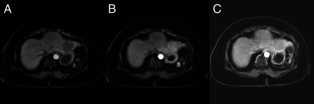
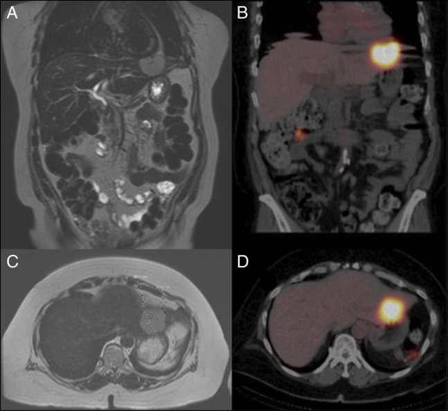
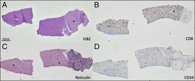
Splenosis is defined as viable splenic tissue that is autotransplanted into other compartments in the body. Intrahepatic splenosis is a rare diagnosis that can be difficult for clinicians to identify. The most common causes of splenosis include abdominal trauma and splenectomy. While most patients with intrahepatic splenosis are asymptomatic, in the presence of risk factors of hepatocellular carcinoma, it is paramount to rule out malignancy. In this report, we present a patient with imaging findings concerning for hepatocellular carcinoma, ultimately diagnosed with percutaneous biopsy and technetium-99m-tagged heat-damaged red blood cell scintigraphy-proven intrahepatic splenosis.
Keywords: hepatocellular carcinoma; intrahepatic splenosis; radiology; splenectomy.
© 2023 The Author(s). Published by Wolters Kluwer Health, Inc. on behalf of The American College of Gastroenterology.
Figures
Similar articles
-
Intrahepatic splenosis: a world review.Clin Exp Hepatol. 2020 Sep;6(3):185-198. doi: 10.5114/ceh.2020.99509. Epub 2020 Sep 30. Clin Exp Hepatol. 2020. PMID: 33145425 Free PMC article. Review.
-
Intrahepatic and intra-abdominal splenosis: A case report and review of literature.World J Hepatol. 2019 Dec 27;11(12):773-779. doi: 10.4254/wjh.v11.i12.773. World J Hepatol. 2019. PMID: 31966909 Free PMC article.
-
Intrahepatic Splenosis: A Rare Case.Curr Med Imaging. 2023;19(6):640-643. doi: 10.2174/1573405619666221212153639. Curr Med Imaging. 2023. PMID: 36515034
-
Masquerading Spleen: A Perplexing Case of Intrahepatic Splenosis Mimicking Hepatocellular Carcinoma.Cureus. 2024 Sep 25;16(9):e70145. doi: 10.7759/cureus.70145. eCollection 2024 Sep. Cureus. 2024. PMID: 39463668 Free PMC article.
-
Management of intrahepatic splenosis:a case report and review of the literature.World J Surg Oncol. 2018 Jun 28;16(1):119. doi: 10.1186/s12957-018-1419-1. World J Surg Oncol. 2018. PMID: 29954390 Free PMC article. Review.
References
-
- Buchbinder J, Lipkoff C. Splenosis: Multiple peritoneal splenic implants following abdominal injury: A report of a case and review of the literature. Surgery. 1939;6:927–34.
-
- Kwok CM, Chen YT, Lin HT, et al. . Portal vein entrance of splenic erythrocytic progenitor cells and local hypoxia of liver, two events cause intrahepatic splenosis. Med Hypotheses. 2006;67:1330–2. - PubMed
-
- Ksiadzyna D, Pena A. Abdominal splenosis. Rev Esp Enferm Dig. 2011;103:421–6. - PubMed
Publication types
LinkOut - more resources
Full Text Sources
