Review
. 2023 Jun 27:10:1199146.
doi: 10.3389/fmed.2023.1199146. eCollection 2023.

Current and potential roles of immuno-PET/-SPECT in CAR T-cell therapy

Affiliations
Review

Current and potential roles of immuno-PET/-SPECT in CAR T-cell therapy

Aditi Mulgaonkar et al. Front Med (Lausanne). .

Abstract

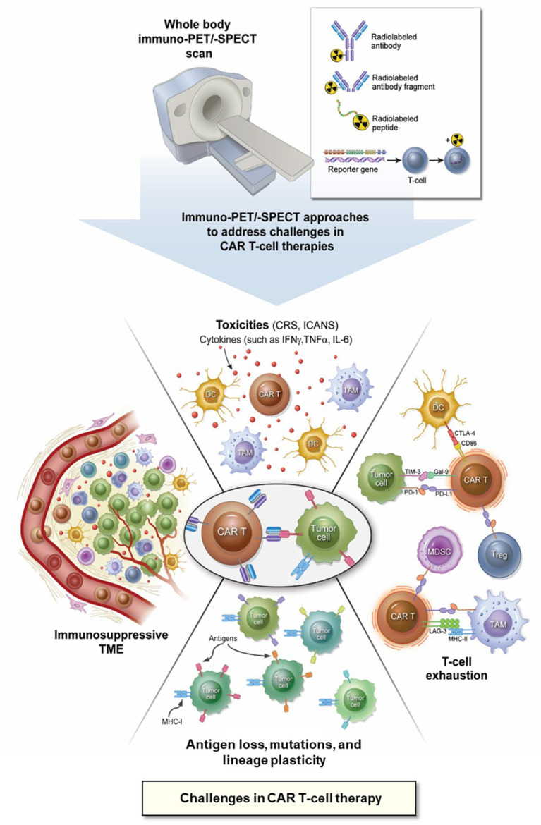
Chimeric antigen receptor (CAR) T-cell therapies have evolved as breakthrough treatment options for the management of hematological malignancies and are also being developed as therapeutics for solid tumors. However, despite the impressive patient responses from CD19-directed CAR T-cell therapies, ~ 40%-60% of these patients' cancers eventually relapse, with variable prognosis. Such relapses may occur due to a combination of molecular resistance mechanisms, including antigen loss or mutations, T-cell exhaustion, and progression of the immunosuppressive tumor microenvironment. This class of therapeutics is also associated with certain unique toxicities, such as cytokine release syndrome, immune effector cell-associated neurotoxicity syndrome, and other "on-target, off-tumor" toxicities, as well as anaphylactic effects. Furthermore, manufacturing limitations and challenges associated with solid tumor infiltration have delayed extensive applications. The molecular imaging modalities of immunological positron emission tomography and single-photon emission computed tomography (immuno-PET/-SPECT) offer a target-specific and highly sensitive, quantitative, non-invasive platform for longitudinal detection of dynamic variations in target antigen expression in the body. Leveraging these imaging strategies as guidance tools for use with CAR T-cell therapies may enable the timely identification of resistance mechanisms and/or toxic events when they occur, permitting effective therapeutic interventions. In addition, the utilization of these approaches in tracking the CAR T-cell pharmacokinetics during product development and optimization may help to assess their efficacy and accordingly to predict treatment outcomes. In this review, we focus on current challenges and potential opportunities in the application of immuno-PET/-SPECT imaging strategies to address the challenges encountered with CAR T-cell therapies.

Keywords: CAR T-cell therapy; cell therapy; immuno-PET; immuno-SPECT; tumor microenvironment.


Conflict of interest statement

The authors declare that the research was conducted in the absence of any commercial or financial relationships that could be construed as a potential conflict of interest.

Figures

Figure 1
Figure 1
Application of immuno-PET/-SPECT imaging approaches to address the challenges with CAR T-cell therapies. CAR, chimeric antigen receptor; CD, cluster of differentiation; CRS, cytokine release syndrome; CTLA-4, cytotoxic T lymphocyte antigen-4; DC, dendritic cell; Gal-9, galectin 9; ICANS, immune effector cell-associated neurotoxicity syndrome; IFNγ, interferon gamma; IL-6, interleukin 6; immuno-PET/-SPECT, immunological positron emission tomography or single-photon emission computed tomography; LAG-3, lymphocyte activation gene 3 protein; MDSC, myeloid-derived suppressor cell; MHC, major histocompatibility complex; PD-1, programmed cell death protein 1; PD-L1, programmed cell death protein ligand 1; TAM, tumor-associated macrophage; TIM-3, T-cell immunoglobulin and mucin domain protein 3; TME, tumor microenvironment; TNFα, tumor necrosis factor alpha, Treg, regulatory T-cell.
Figure 2
Figure 2
Immuno-PET imaging with [89Zr]Zr-Rituximab in patients with DLBCL treated with a therapeutic dose of rituximab. (A) Immuno-PET imaging shows intense tumor uptake (top panel) concordant with CD20-positive IHC staining of the inguinal lymph node biopsy sample (B, top panel). Corresponding [18F]FDG-PET images are shown in the bottom panel (A). (C) CD20-negative tumor shows no appreciable uptake of [89Zr]Zr-Rituximab (top panel) concordant with IHC staining of the biopsy (B, bottom panel). In contrast, the tumor exhibits a focal spot on [18F]FDG-PET images (C, bottom panel). Shown in (A) and (C) are attenuation-corrected PET, low-dose CT, and fused PET/CT images from left to right. Reproduced with slight format modifications from the open-access article in ref. (81) under the Creative Commons license (http://creativecommons.org/licenses/by/4.0/).
Figure 3
Figure 3
Immuno-SPECT/CT imaging of HCC1954 HER2+ tumor-bearing mice injected with [67Cu]Cu-NOTA-Pertuzumab. (A) Representative maximum intensity projection (MIP) of SPECT/CT images in mouse groups as indicated at days 2 and 5 post-treatment (yellow arrows indicate the tumors). (B) Actual radioactivity concentration in tumors (MBq/ml) on days 2 and 5 (without decay correction). Reproduced from the open access article in ref. (42) under the Creative Commons license (http://creativecommons.org/licenses/by/4.0/).
Figure 4
Figure 4
Integrated immuno-PET, IHC, and hematoxylin and eosin (H&E) images of mouse tumorgraft lines from corresponding patient tumors with high and low PD- L1 expression groups. (Top) Schematic representative of the workflow. (Middle) Representative whole-body MIP immuno-PET images (posterior view) of mice, one from each group. The corresponding PD-L1 expression ranges measured by IHC and the volume of the tumor in the mouse as indicated are shown below (n = 3–4 for each line; a single remaining XP258 mouse is not included). Tumors are indicated with a yellow lasso. (Bottom) PD-L1 IHC and H&E staining of the corresponding tumor tissues explanted from the TG models. Patient tumor samples shown as a reference. A part of this figure has been reproduced from the referenced article (41) under reuse permission from standard copyright in AACR journals for authors.
Figure 5
Figure 5
Whole-body immuno-PET imaging with [89Zr]Zr-IAB22M2C in a patient at 24 h post-injection. (A) Intense uptake is noted in lymph nodes. (B, C) Fusion image at 24 h shows [89Zr]Zr-IAB22M2C uptake in lesion and deltoid (B), which were also [18F]FDG positive (C). (D) H&E stained section shows melanoma tumor nodules on the right within skeletal muscle. (E) IHC highlights the presence of CD8+ T-cells at the periphery and infiltrating tumors [reproduced with permission from original publication Pandit-Taskar et al. (183)].
Figure 6
Figure 6
Representative [18F]FB-IL2 PET images of human melanoma. (A) Transversal PET/CT image of three regions showing high [18F]FB-IL2 uptake. (B) MIP image of the same patient showing multiple areas of high radiotracer accumulation in the lungs. Reproduced from the open access article in ref. (158) under the Creative Commons license (http://creativecommons.org/licenses/by/4.0/).

Similar articles

Cited by

References

    1. Martínez-Lostao L, Anel A, Pardo J. How do cytotoxic lymphocytes kill cancer cells? Clin Cancer Res. (2015) 21:5047–56. 10.1158/1078-0432.CCR-15-0685 - DOI - PubMed
    1. Zhou J, Shen X, Huang J, Hodes RJ, Rosenberg SA, Robbins PF. Telomere length of transferred lymphocytes correlates with in vivo persistence and tumor regression in melanoma patients receiving cell transfer therapy. J Immunol. (2005) 175:7046–52. 10.4049/jimmunol.175.10.7046 - DOI - PMC - PubMed
    1. Robbins PF, Lu YC, El-Gamil M, Li YF, Gross C, Gartner J, et al. . Mining exomic sequencing data to identify mutated antigens recognized by adoptively transferred tumor-reactive T cells. Nat Med. (2013) 19:747–52. 10.1038/nm.3161 - DOI - PMC - PubMed
    1. Rosenberg SA. IL-2: the first effective immunotherapy for human cancer. J Immunol. (2014) 192:5451–8. 10.4049/jimmunol.1490019 - DOI - PMC - PubMed
    1. Rosenberg SA, Yang JC, Sherry RM, Kammula US, Hughes MS, Phan GQ, et al. . Durable complete responses in heavily pretreated patients with metastatic melanoma using T-cell transfer immunotherapy. Clin Cancer Res. (2011) 17:4550–7. 10.1158/1078-0432.CCR-11-0116 - DOI - PMC - PubMed

LinkOut - more resources