Review
. 2023 Jun 27:10:1009411.
doi: 10.3389/fcvm.2023.1009411. eCollection 2023.

Tissue characterization of benign cardiac tumors by cardiac magnetic resonance imaging, a review of core imaging protocol and benign cardiac tumors

Affiliations
Review

Tissue characterization of benign cardiac tumors by cardiac magnetic resonance imaging, a review of core imaging protocol and benign cardiac tumors

Imran Haider et al. Front Cardiovasc Med. .

Abstract

Generally, cardiac masses are initially suspected on routine echocardiography. Cardiac magnetic resonance (CMR) imaging is further performed to differentiate tumors from pseudo-tumors and to characterize the cardiac masses based on their appearance on T1/T2-weighted images, detection of perfusion and demonstration of gadolinium-based contrast agent uptake on early and late gadolinium enhancement images. Further evaluation of cardiac masses by CMR is critical because unnecessary surgery can be avoided by better tissue characterization. Different cardiac tissues have different T1 and T2 relaxation times, principally owing to different internal biochemical environments surrounding the protons. In CMR, the signal intensity from a particular tissue depends on its T1 and T2 relaxation times and its proton density. CMR uses this principle to differentiate between various tissue types by weighting images based on their T1 or T2 relaxation times. Generally, tumor cells are larger, edematous, and have associated inflammatory reactions. Higher free water content of the neoplastic cells and other changes in tissue composition lead to prolonged T1/T2 relaxation times and thus an inherent contrast between tumors and normal tissue exists. Overall, these biochemical changes create an environment where different cardiac masses produce different signal intensity on their T1- weighted and T2- weighted images that help to discriminate between them. In this review article, we have provided a detailed description of the core CMR imaging protocol for evaluation of cardiac masses. We have also discussed the basic features of benign cardiac tumors as well as the role of CMR in evaluation and further tissue characterization of these tumors.

Keywords: atrial myxoma; benign cardiac tumors; cardiac MRI (CMR); fibroelastoma; magnetic resonance imaging; tissue characterization.


Conflict of interest statement

The authors declare that the research was conducted in the absence of any commercial or financial relationships that could be construed as a potential conflict of interest.

Figures

Figure 1
Figure 1
Most common locations of cardiac tumors.
Figure 2
Figure 2
A core imaging protocol for cardiac mass evaluation. FSE, ast spin echo; IR, inversion recovery; MOLLI, modified look locker inversion recovery; SSFP, steady state free procession; SPIR, saturation spectral pre-saturation inversion recovery; TI, inversion time.
Figure 3
Figure 3
A 51-year-old female with left atrial myxoma. (A) Absence of perfusion of the lesion on first pass perfusion images (red arrow). (B) Following gadolinium administration, there is brisk, heterogeneous enhancement of this lesion on late gadolinium enhanced images (red arrow).
Figure 4
Figure 4
A 69-year-old female with right ventricular apical myxoma. (A) Localizer CMR images showing a small hyperintense mass in the distal RV (red arrow). (B) The mass showing high signal intensity on axial T2weighted, dark blood, double inversion recovery fast spin echo images (red arrow). (C) The mass shows absence of perfusion on first pass perfusion images (red arrow). (D) Heterogeneous enhancement of this lesion is noted on late gadolinium enhanced images. (E) There are small central areas of non-enhancement representing necrotic or hemorrhagic material (yellow arrow). RV, right ventricle.
Figure 5
Figure 5
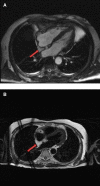
A 53-year-old male with lipomatous hypertrophy of the intra-atrial septum. (A) Cine CMR images showing diffuse enlargement and fatty replacement of the intra-atrial septum, consistent with lipomatous hypertrophy of the intra-atrial septum (red arrow). (B) Characteristic “dumbbell shaped” appearance of the mass on axial dark blood, double inversion recovery fast spin echo images (red arrow).
Figure 6
Figure 6
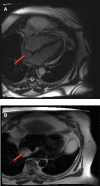
A 61-year-old male with lipomatous hypertrophy of the intra-atrial septum. (A) Cine CMR images showing diffuse enlargement and fatty replacement of the intra-atrial septum, consistent with lipomatous hypertrophy of the intra-atrial septum (red arrow). (B) Characteristic “dumbbell shaped” appearance of the mass on axial dark blood, double inversion recovery fast spin echo images (red arrow).
Figure 7
Figure 7
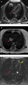
A 68-year-old female with intra-atrial septal lipoma. (A) (On line video 3), CMR cine images showing a sharply marginated mass arising from the intraatrial septum (red arrow). (B) This mass has same signal intensity (high) as surrounding fat on axial T1-weighted, dark blood, double inversion recovery fast spin echo images (red arrow). (C) Fat suppression sequence representing suppression of surrounding fat (white arrow) as well as the mass in the intra-atrial septum (red arrow).
Figure 8
Figure 8
A 50-year-old female with intra-atrial septal lipoma. (A) This mass has signal intensity (high) as surrounding fat on axial T2-weighted, dark blood, double inversion recovery fast spin echo images (red arrow). (B) This mass is suppressed on axial triple inversion recovery images (red arrow). (C) Complete absence of contrast agent uptake on late gadolinium enhanced images (red arrow).
Figure 9
Figure 9
A 71-year-old female with aortic valve fibroelastoma. (A) This mass showing hyper-intense signal on T2weighted, dark blood, double inversion recovery fast spin echo images (red arrow).
Figure 10
Figure 10
A 31-year-old male with left ventricular fibroma. (A) This mass demonstrates no perfusion on first-pass imaging (red arrow, short axis view). (B–F) Homogeneous intense enhancement of the mass is noted on late gadolinium enhanced images (red arrows, 3chamber and short axis views).
Figure 11
Figure 11
A 54-year-old male with right ventricular pheochromocytoma. (A) The mass is isointense on T1- weighted, dark blood, double inversion recovery fast spin echo images (red arrow). (B) The mass demonstrates peripheral hyper-vascularity with central areas of nonenhancement on first pass perfusion images (red arrow). (C) There is central enhancement of this mass on late gadolinium enhanced images (red arrow).
Figure 12
Figure 12
A 53-year-old male with inter-atrial septal pheochromocytoma. (A) The mass is bright on T2- weighted, dark blood, double inversion recovery fast spin echo images (red arrow). (B) This mass shows heterogeneous enhancement on late gadolinium enhanced images (red arrow).
Figure 13
Figure 13
Cardiac Hemagioma in a 40 years old male. In Right ventricle there is round contrast enhancing lesion, involving the right ventricular cavity shown in Figure A is cardiac hemangioma.
Figure 14
Figure 14
A mature teratoma in a 16 years old female patient. (A). Contrast enhancing CT chest showing a calcified, fatty lesion inside heart, calcifications are multifocal representing huge mature cardiac teratoma.

References

    1. Sparrow PJ, Kurian JB, Jones TR, et al. MR Imaging of cardiac tumors. Radiographic. (2005) 25:1255–76. 10.1148/rg.255045721 - DOI - PubMed
    1. Sütsch G, Jenni R, von Segesser L, Schneider J. Heart tumors: incidence, distribution, diagnosis exemplified by 20,305 echocardiographies. Schweiz Med Wochenschr. (1991) 121:621–29. PMID: 2047823 - PubMed
    1. Bruce CJ. Cardiac tumors: diagnosis and management. Heart. (2011) 97:151–60. 10.1136/hrt.2009.186320 - DOI - PubMed
    1. Elbardissi AW, Dearani JA, Daly RC, et al. Survival after resection of primary cardiac tumors: a 48-year experience. Circulation. (2008) 118:S7–15. 10.1161/CIRCULATIONAHA.107.783126 - DOI - PubMed
    1. Roberts WC. Primary and secondary neoplasms of the heart. Am J Cardiol. (1997) 80:671–82. 10.1016/S0002-9149(97)00587-0 - DOI - PubMed

LinkOut - more resources