. 2023 Oct:301:122238.
doi: 10.1016/j.biomaterials.2023.122238. Epub 2023 Jul 5.

Bioactive silica nanoparticles target autophagy, NF-κB, and MAPK pathways to inhibit osteoclastogenesis

Affiliations

Bioactive silica nanoparticles target autophagy, NF-κB, and MAPK pathways to inhibit osteoclastogenesis

Jamie Arnst et al. Biomaterials. 2023 Oct.

Abstract

Spherical 50 nm silica-based nanoparticles (SiNPs) promote healthy bone homeostasis and maintenance by supporting bone forming osteoblast lineage cells while simultaneously inhibiting the differentiation of bone resorbing osteoclasts. Previous work demonstrated that an intraperitoneal injection of SiNPs in healthy mice - both young and old - increased bone density and quality, suggesting the possibility that SiNPs represent a dual action therapeutic. However, the underlying mechanisms governing the osteoclast response to SiNPs have yet to be fully explored and defined. Therefore, the goals of this study were to investigate the cellular and molecular mechanisms by which SiNPs inhibit osteoclastogenesis. SiNPs strongly inhibited RANKL-induced osteoclast differentiation within the first hours and concomitantly inhibited early transcriptional regulators such as Nfatc1. SiNPs simultaneously stimulated expression of autophagy related genes p62 and LC3β dependent on ERK1/2 signaling pathway. Intriguingly, SiNPs were found to stimulate autophagosome formation while inhibiting the autophagic flux necessary for RANKL-stimulated osteoclast differentiation, resulting in the inhibition of both the canonical and non-canonical NF-κB signaling pathways and stabilizing TRAF3. These results suggest a model in which SiNPs inhibit osteoclastogenesis by inhibiting the autophagic machinery and RANKL-dependent functionality. This mechanism of action defines a novel therapeutic strategy for inhibiting osteoclastogenesis.

Keywords: Autophagy; Bone resorption; LC3β; Nfatc1; Osteoporosis; p62.


Conflict of interest statement

Declaration of competing interest The authors declare that they have no known competing financial interests or personal relationships that could have appeared to influence the work reported in this paper.

Figures

Fig.1:
Fig.1:
Silica Nanoparticles inhibit RANKL induced gene expression during osteoclast differentiation. RAW264.7 cells were treated with SiNPs (25μg/mL) for 30 minutes prior to addition of RANKL (15ng/ml) for indicated times and cells harvested for RNA and analysis of gene expression. (a) Osteoclast transcriptional regulators, (b) Osteoclast differentiation marker genes, (c) autophagy genes, and (d) inhibitory transcription factors. RNA was analyzed by qRT-PCR and fold change calculated using ΔΔCT method. Data expressed as Mean ±SEM. Samples run in triplicate. Representative of multiple experiments. (e) Western blot of RAW264.7 cells treated as in (a) and probed with antibodies as indicated. (f) Raw264.7 cells were untreated or treated with RANKL and NPs as in (a) and a live/dead assay performed (n=3). Results are presented as percent change from untreated cells. *P<0.05, ***P<0.0005, relative to corresponding +RANKL control by student’s t test.
Fig. 2:
Fig. 2:
SiNP inhibition of osteoclastogenesis is time dependent. (a) RAW264.7 cells were treated with RANKL (15ng/mL) and SiNPs (25μg/mL) added at times indicated. After 3 days, osteoclastogenesis was analyzed by TRAP staining. (b) RAW cells were treated as in (a) 4, 8, 16 h after RANKL addition and cells harvested for RNA at 24h and Nfatc1 quantified by qRT-PCR. (c) Fluorescent SiNPs were added to RAW264.7 cells and fluorescence microscopy was used to track and quantify internalization by RFI (Relative Fluorescence Intensity) (20x) (d), over time. (e) SiNPs were added to RAW264.7 cells for 2h, removed by rinsing with medium, followed by treatment with RANKL (15ng/mL) for 24h and harvested for RNA analysis. RNA was analyzed by qRT-PCR for genes as indicted and fold change calculated using ΔΔCT method. Data are expressed as Mean ±SD. Samples run in triplicate. Representative of multiple experiments. #P<0.05, ##P<0.005, ###P<0.0005 compared to untreated, *P<0.05, **P<0.005, ***P<0.0005 relative to RANKL by student’s t test. &&&&P<0.0001 by One way ANOVA.
Fig. 3:
Fig. 3:
ERK1/2 signaling is required for RANKL induced Nfatc1 expression and SiNPs induced p62 expression. (a) RAW264.7 cells were treated with RANKL (15ng/ml) in the absence or presence of SiNPs (25μg/ml) and cells were harvested for Western blotting (whole cell lysate) at indicated times and probed with antibodies as indicated. (b) RAW264.7 cells were pretreated with U0126 at indicated concentrations prior to addition of RANKL (15ng/ml) in the absence or presence of SiNPs (25μg/ml) for 6hr and harvested RNA analyzed by qRT-PCR for Nfatc1 and (c) p62. (d) Cells were treated as above and harvested after 6h for Western blotting (whole cell) and probed with antibodies as indicated. Data are expressed as Mean ±SD. Samples run in triplicate. Representative of multiple experiments. #P<0.05, ##P<0.005, ###P<0.0005 compared to untreated, *P<0.05, **P<0.005, ***P<0.0005 relative to RANKL treated, and &P<0.05, &&P<0.005, &&&P<0.0005 relative to RANKL+SiNP by student’s t test.
Fig. 4:
Fig. 4:
SiNPs inhibit RANKL induced AP-1 and NF-κB signaling. (a) RAW264.7 cells were treated with RANKL (15ng/ml) in the absence or presence of SiNPs (25μg/ml) for indicated times and whole cell lysate harvested for Western blotting of JNK pathway. (b) Cells were treated with RANKL and SiNPs as in (a) for indicated times and harvested for nuclear lysate and analyzed by Western blotting for AP-1 proteins. Representative of multiple experiments. (c) Cell lysate as in (b) and whole cell lysate was probed for NF-kB pathway proteins. (d) Cells were treated as in (a) for indicated times and harvested for nuclear lysate and analyzed by Western blotting for NF-κB proteins. Representative of multiple experiments.
Fig. 5:
Fig. 5:
SiNPs stabilize TRAF3 protein levels after RANKL treatment. (a) RAW264.7 cells were treated with RANKL (15ng/ml) in the absence or presence of SiNPs (25μg/ml) for indicated times and whole cell lysate analyzed by Western blotting at indicated times. (b) Cells were treated as in (a) and whole cell lysate analyzed by Western blotting. Representative of multiple experiments. (c) Cells were stained by immunohistochemistry for TRAF3 and nuclei stained with Hoescht at indicated times after addition of RANKL (15ng/ml) and +/− SiNPs (25μg/ml) (scale bar=10μm). (d) TRAF3 RFI were quantified (50 cells/condition) by ImageJ. *P<0.05, **P<0.005 relative to RANKL by student’s t test.
Fig. 6:
Fig. 6:
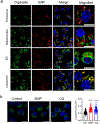
SiNPs co-localize with the specific organelles. (a) RAW264.7 cells were treated with SiNPs (25μg/ml) and cells live imaged by confocal microcopy. SiNPs (red), Lysosome (Lyso-Tracker), Mitochondria (Mito-Tracker), Endosome (Tfn-FITC) or Endoplasmic Reticulum (ER-Tracker) (green) and Nuclei were stained with Hoescht 33342 (blue). Scale bars=10μm, Magnified Scale bars=5μm. (b) RAW264.7 cells were treated for 4h as indicated and were incubated with LysoTracker (green) and Hoescht 3342 (blue) before imaging as in (a). Scale bars=10μm. Quantification of the lysosomal size per cell from the immunofluorescence images are shown (50 cells/condition). ****P<0.0001 relative to control by student’s t test.
Fig. 7:
Fig. 7:
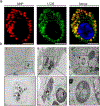
SiNPs co-localize and are found in the Autophagosome/Autolysosome like structures. (a) RAW264.7 cells were treated with SiNPs (red) (25μg/ml) and fixed after 4h autophagosomes visualized with LC3β antibody (green). Nuclei were stained with Hoescht 33342 (blue). Scale bar=5μm. Representative of multiple experiments. (b) RAW264.7 cells were treated with metal core nanoparticles for 24h (i,ii) or control (iii) and visualized by Transmission electron Microscopy. Scale bars (200nm (i, iii) or 500nm (ii)) and insets (50nm (i, iii) or 100nm (ii)).
Fig. 8:
Fig. 8:
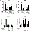
Inhibition of autophagy blocks RANKL induced gene expression. (a) RAW264.7 cells were treated with RANKL (15ng/ml) and autophagy inhibitors 3-MA (500μM) or CQ (2.5μM) at indicated times after RANKL. (b) RAW264.7 cells were treated with RANKL and SiNPs (25μg/ml) and autophagy inhibitors as indicated 3MA (500μM) and (10μM) CQ, and RNA harvested after 16h. RNA was analyzed by qRT-PCR and fold change calculated using ΔΔCT method. Data are expressed as Mean ±SD. Samples run in triplicate. Representative of multiple experiments. #P<0.05, ##P<0.005, ###P<0.0005 compared to untreated, *P<0.05, **P<0.005, ***P<0.0005 relative to RANKL by student’s t test.
Fig. 9:
Fig. 9:
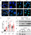
SiNPs stimulate autophagosome formation and directly interact with autophagosomal proteins. (a) RAW264.7 cells were treated with RANKL (15ng/ml) or RANKL+SiNPs (25μg/ml) for times indicated and autophagosomes visualized with LC3β antibody (green) and nuclei with DAPI (blue). Scale bar=10μm. (b) Puncta were counted and averaged (40 cells/condition/2 exp.). *P<0.05, ****P<0.00005 relative to RANKL and #P<0.05, ###P<0.0005, ####P<0.00005 relative to previous time point within condition by One-way ANOVA with Sidak’s multiple comparisons test. (c) RAW264.7 cells were treated with SiNPs (25μg/ml) followed by RANKL (15ng/ml) and whole cell lysate analyzed by Western blotting at indicated time. (d) Magnetic core SiNPs (with OH or PEGlyated surface) were used to pull-down associated proteins from RAW264.7 cells or FBS (negative control). Whole cell lysate (WCL) prior to pulldown is shown for comparison. After washing the associated proteins were analyzed by Western blotting.
Fig. 10:
Fig. 10:
SiNPs stimulate autophagosome formation but do not alter autophagic flux. (a) RAW264.7 cells were stably transfected with an autophagic flux probe (GFP-LC3-RFP-LC3ΔG) and treated with RANKL (RL) (15ng/ml), SiNPs (25μg/ml), Chloroquine (CQ) (10uM) or a combination as indicated and for times indicated and visualized by microscopy. Scale bar=20μm. (b) Total GFP/RFD fluorescence was quantified for the 6h timepoint, and ratio calculated and plotted as a percent relative to control (untreated) cells (50 cells/condition). *P<0.05, ****P<0.0001 as indicated and &P<0.05 relative to untreated by student’s t test. (c) RAW264.7 cells were treated as in (a) with SiNPs for 1h and imaged.
Fig. 11:
Fig. 11:
Working model of the effects of SiNPs on RANKL induced osteoclast differentiation. The internalization of SiNPs stimulated phosphorylation and activation of the ERK1/2 signaling pathway and increased expression of autophagy genes p62 and LC3β. SiNPs strongly stimulate autophagy and localize to autophagosomes and autolysosomes while simultaneously inhibiting autophagic flux (dashed red line) which is required for RANKL induced signaling of the AP-1 and NF-κB transcriptional regulators to stimulate osteoclastogenesis. The blocked degradation of autolysosome components results in the inhibition of TRAF3 degradation (stabilization) thereby inhibiting downstream RANKL signaling.

References

    1. Beck GR Jr., Ha SW, Camalier CE, Yamaguchi M, Li Y, Lee JK, Weitzmann MN, Bioactive silicabased nanoparticles stimulate bone-forming osteoblasts, suppress bone-resorbing osteoclasts, and enhance bone mineral density in vivo, Nanomedicine: nanotechnology, biology, and medicine 8(6) (2012) 793–803. - PMC - PubMed
    1. Ha SW, Viggeswarapu M, Habib MM, Beck GR Jr., Bioactive effects of silica nanoparticles on bone cells are size, surface, and composition dependent, Acta Biomater 82 (2018) 184–196. - PMC - PubMed
    1. Ha SW, Camalier CE, Beck GR Jr., Lee JK, New method to prepare very stable and biocompatible fluorescent silica nanoparticles, Chem Commun (Camb) (20) (2009) 2881–3. - PMC - PubMed
    1. Ha S-W, Camalier CE, Weitzmann MN, Beck GR, Lee J-K, Long-Term Monitoring of the Physicochemical Properties of Silica-Based Nanoparticles on the Rate of Endocytosis and Exocytosis and Consequences of Cell Division, Soft Materials 11(2) (2013) 195–203. - PMC - PubMed
    1. Udagawa N, Takahashi N, Akatsu T, Tanaka H, Sasaki T, Nishihara T, Koga T, Martin TJ, Suda T, Origin of osteoclasts: mature monocytes and macrophages are capable of differentiating into osteoclasts under a suitable microenvironment prepared by bone marrow-derived stromal cells, Proc Natl Acad Sci U S A 87(18) (1990) 7260–4. - PMC - PubMed

Publication types