. 2023 Oct:76:101772.
doi: 10.1016/j.molmet.2023.101772. Epub 2023 Jul 11.

EndoC-βH5 cells are storable and ready-to-use human pancreatic beta cells with physiological insulin secretion

Affiliations

EndoC-βH5 cells are storable and ready-to-use human pancreatic beta cells with physiological insulin secretion

Bruno Blanchi et al. Mol Metab. 2023 Oct.

Abstract

Objectives: Readily accessible human pancreatic beta cells that are functionally close to primary adult beta cells are a crucial model to better understand human beta cell physiology and develop new treatments for diabetes. We here report the characterization of EndoC-βH5 cells, the latest in the EndoC-βH cell family.

Methods: EndoC-βH5 cells were generated by integrative gene transfer of immortalizing transgenes hTERT and SV40 large T along with Herpes Simplex Virus-1 thymidine kinase into human fetal pancreas. Immortalizing transgenes were removed after amplification using CRE activation and remaining non-excized cells eliminated using ganciclovir. Resulting cells were distributed as ready to use EndoC-βH5 cells. We performed transcriptome, immunological and extensive functional assays.

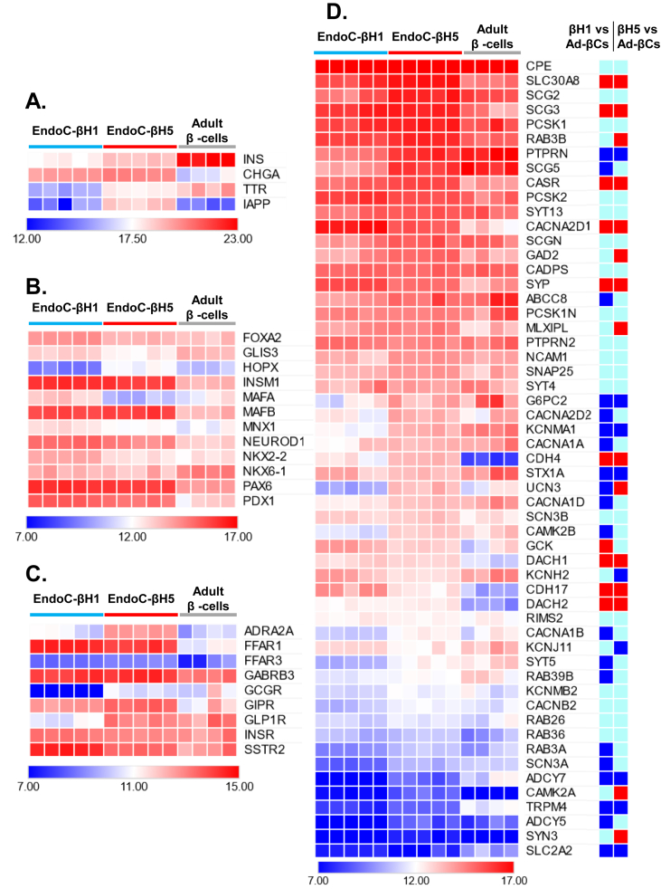
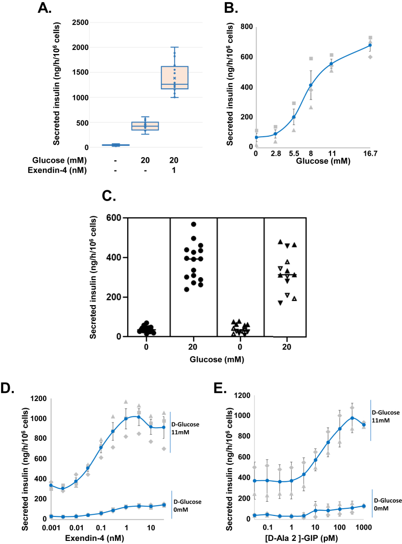
Results: Ready to use EndoC-βH5 cells display highly efficient glucose dependent insulin secretion. A robust 10-fold insulin secretion index was observed and reproduced in four independent laboratories across Europe. EndoC-βH5 cells secrete insulin in a dynamic manner in response to glucose and secretion is further potentiated by GIP and GLP-1 analogs. RNA-seq confirmed abundant expression of beta cell transcription factors and functional markers, including incretin receptors. Cytokines induce a gene expression signature of inflammatory pathways and antigen processing and presentation. Finally, modified HLA-A2 expressing EndoC-βH5 cells elicit specific A2-alloreactive CD8 T cell activation.

Conclusions: EndoC-βH5 cells represent a unique storable and ready to use human pancreatic beta cell model with highly robust and reproducible features. Such cells are thus relevant for the study of beta cell function, screening and validation of new drugs, and development of disease models.

Keywords: Glucose and incretin stimulated insulin secretion; Human beta cell function; Human pancreatic beta cell line; Type-I diabetes disease model.


Conflict of interest statement

Declaration of Competing Interest BB, MT, AP, CC, MP, AB and HO are or were employees at Human Cell Design SA, France, the company that commercializes EndoC-βH1 and EndoC-βh5 cells and associated media. RS, PC and PR are shareholders at HCD.

Figures

Figure 1
Figure 1
Production of ready to use EndoC-βH5 cells and characterization by immuno-labeling. (A) Schematic representation of integrated lentiviral vector used to produce EndoC-βH5 cells. Upon integration the loxP site located into the deleted U3 3′ LTR region is duplicated. The entire integrated sequences are flanked with loxP sites thus allowing CRE mediated excision. The integrated sequence contains a first transcription unit expressing SV40 LT or hTERT under the control of the rat insulin promoter (RIP) and a second unit expressing HSV1 TK under the control of the polyomavirus enhancer (pY). (B) The immortalized cell line is transduced with a lentiviral vector expressing a tamoxifen inducible form of CRE (CRE-ERT2) expressed under the control of RIP. After massive amplification, cells are treated for 7 days with 1 μM tamoxifen followed by 1 day of ganciclovir to allow SV40LT and hTERT excision and destruction of non-excised cells that expressed TK. The resulting EndoC-βH5 cells are frozen to produce ready to use vials. Panels A and B were created with BioRender.com. (C) FACS analysis profile (black curves) of insulin, PDX1 and NKX6.1 expression in EndoC-βH5 cells. The grey curve represents the isotype control. (D) Phase contrast photograph of ready to use EndoC-βH5 thawed cells. (E) Insulin (red), PDX1 (green) and nuclei (DAPI, blue) immunofluorescent staining of EndoC-βH5 cells. Scale bar = 50 μm.
Figure 2
Figure 2
Heat map of gene expression profiling in EndoC-βH5, EndoC-βH1 and FACS purified human adult beta cells. The heat maps show the variance stabilizing transformation (VST) of the count data sets normalized to library size of 5 independent EndoC-βH5 and EndoC-βH1 cell preparations and 4 publicly available data sets of FACS purified human adult beta cells (adult β cells). (A) INS, CHGA, TTR and IAPP represent the top four non-mitochondrial transcripts expressed in EndoC-βH5 cells, ranking 1st, 7th, 14th and 18th respectively. (B) List of transcription factors essential for beta cell development and identity. (C) List of receptors regulating insulin secretion in beta cells. (D) Gene lists of transcripts important for beta cell function, generated using published transcriptomics data, literature data, and manual curation. Genes do not overlap with the 3 lists presented in A, B and C. Left panel, heat maps of expression (VST) in EndoC-βH1, EndoC-βH5 and adult β cells. Right panel, differential expression between EndoC-βH1 and adult β cells and EndoC-βH5 and adult β cells. Significant changes corresponding to a fold change >2 and an FDR <0.05 are represented in blue and red for reduced or increased expression respectively. Light green squares represent non-significant changes.
Figure 3
Figure 3
Robust and highly reproducible glucose and GLP-1/GIP receptor agonist stimulated insulin secretion by EndoC-βH5 cells, Box-plot (A) and graphs (B, C, D) showing static insulin secretion by EndoC-βH5 cells subjected to various stimuli and expressed as ng insulin that is secreted per h and per million cells (ng/h/106 cells). (A) Box-plot of GSIS results from 16 independent EndoC-βH5 batches. Cells were stimulated with d-Glucose (20 mM) or d-Glucose (20 mM) plus exendin-4 (1 nM). (B)d-glucose dose response (2.8–16.7 mM). Graph shows results of 3 independent experiments (grey symbols) and average (blue circles and curve). (C) Plot of individual GSIS results (LG, 0 mM; HG, 20 mM) obtained at HCD (left, circles, 16 independent experiments) and in four European laboratories (right, triangles, 13 independent experiments). Upward/downward and solid/empty triangles correspond each of the four separate laboratories. (D) Exendin-4 dose response (0.001–33 nM) in presence (top) or absence (bottom) of d-Glucose (11 mM). Graph shows results from 3 independent experiments (grey symbols) and average (blue circles and curve). Bottom experiment (no d-Glucose) was repeated twice. (E) [D-Ala2]-GIP dose response (0.033 pM to 1 nM) in presence (top) or absence (bottom) of d-Glucose (11 mM). Graph shows results from 3 independent experiments (grey symbols) and average (blue circles and curve).
Figure 4
Figure 4
Dynamic insulin secretion by EndoC-βH5 spheres. (A) Morphology of EndoC-βH5 spheres. (B) Dynamic insulin secretion by EndoC-βH5 spheres in a perifusion setting in presence of 1.5 mM glucose (G1.5), 8.5 mM glucose (G8.5) or 40 mM KCl (KCl). Five experiments were performed, using two independent preparations of spheres at three (day 11, 18 and 25) and two (day 13 and 20) time points. Insulin secretion is expressed as ng insulin secreted per time-point (2-minute) and per million cells (ng/106 cells). (C) Dynamic insulin secretion by EndoC-βH5 spheres maintained in 0.5 mM glucose showing initial first-phase peak. Spheres were stimulated with 3 mM glucose (G3), 17 mM glucose (G17) or 40 mM KCl (KCl). In two of the experiments, G17 and second G3 incubation duration were longer. This additional time is not shown (broken X axis) Five experiments were performed, using two independent preparations of spheres. Insulin release is expressed as fold induction relative to the first G3 point.
Figure 5
Figure 5
EndoC-βH5 are responsive to cytokines and EndoC-βH5/A2 is applicable in T cell reactivity assays. (A) Volcano plot of protein coding transcripts expression detected in RNA-seq of EndoC-βH5 cells treated with IFNγ/IL1β for 24 h (n = 3). The Volcano plot was generated using EnhancedVolcano. (B) Dot plot of enriched gene ontology terms within the group of transcripts upregulated by cytokine treatment (logFC>2, FDR<0.05). The dot plot was generated using clusterProfiler. (C) Heatmap of the most upregulated transcripts (logFC>5, FDR< 0.05). Read counts were averaged over the triplicates and only transcripts with an average > 3 are depicted. The heatmap was generated using Pheatmap. (D) HLA-A2 expression in EndoC-βH5 and EndoC-βH5/HLA-A2 cells in normal or IFNγ/IL1β treated condition. Data are presented as mean fluorescent intensity (MFI). (E) MIP-1β secretion of allo-HLA-A2 reactive CTLs after overnight coculture with EndoC-βΗ5 (left panel) or EndoC-βΗ5/HLA-A2 (right panel). Target cells were untreated or treated with IFNγ/IL1β with or without 4 μΜ baricitinib (Bari) for 24 h. N = 3 independent experiments. N.B. In this figure, cytokines means IFNγ and IL1β.

References

    1. Diseases G.B.D., Injuries C. Global burden of 369 diseases and injuries in 204 countries and territories, 1990-2019: a systematic analysis for the Global Burden of Disease Study 2019. Lancet. 2020;396(10258):1204–1222. Epub 2020/10/19. - PMC - PubMed
    1. Singer M.E., Dorrance K.A., Oxenreiter M.M., Yan K.R., Close K.L. The type 2 diabetes 'modern preventable pandemic' and replicable lessons from the COVID-19 crisis. Prev Med Rep. 2022;25:101636. Epub 2021/12/16. - PMC - PubMed
    1. Ravassard P., Hazhouz Y., Pechberty S., Bricout-Neveu E., Armanet M., Czernichow P., et al. A genetically engineered human pancreatic beta cell line exhibiting glucose-inducible insulin secretion. J Clin Invest. 2011;121(9):3589–3597. Epub 2011/08/26. - PMC - PubMed
    1. Benazra M., Lecomte M.J., Colace C., Muller A., Machado C., Pechberty S., et al. A human beta cell line with drug inducible excision of immortalizing transgenes. Mol Metabol. 2015;4(12):916–925. Epub 2016/02/26. - PMC - PubMed
    1. Scharfmann R., Pechberty S., Hazhouz Y., von Bulow M., Bricout-Neveu E., Grenier-Godard M., et al. Development of a conditionally immortalized human pancreatic beta cell line. J Clin Invest. 2014;124(5):2087–2098. Epub 2014/03/29. - PMC - PubMed

Publication types