. 2023 Sep 28;62(3):2202210.
doi: 10.1183/13993003.02210-2022. Print 2023 Sep.

Oxidised IL-33 drives COPD epithelial pathogenesis via ST2-independent RAGE/EGFR signalling complex

Affiliations

Oxidised IL-33 drives COPD epithelial pathogenesis via ST2-independent RAGE/EGFR signalling complex

Sam Strickson et al. Eur Respir J. .

Abstract

Background: Epithelial damage, repair and remodelling are critical features of chronic airway diseases including chronic obstructive pulmonary disease (COPD). Interleukin (IL)-33 released from damaged airway epithelia causes inflammation via its receptor, serum stimulation-2 (ST2). Oxidation of IL-33 to a non-ST2-binding form (IL-33ox) is thought to limit its activity. We investigated whether IL-33ox has functional activities that are independent of ST2 in the airway epithelium.

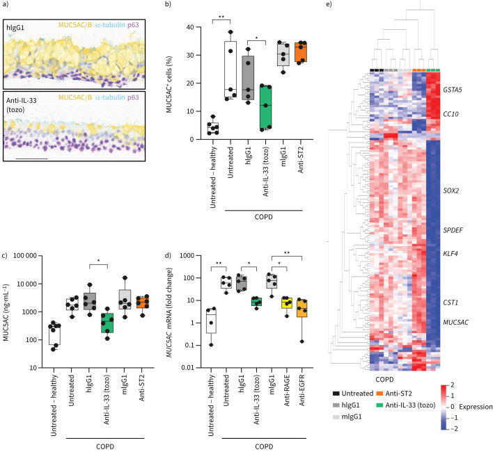
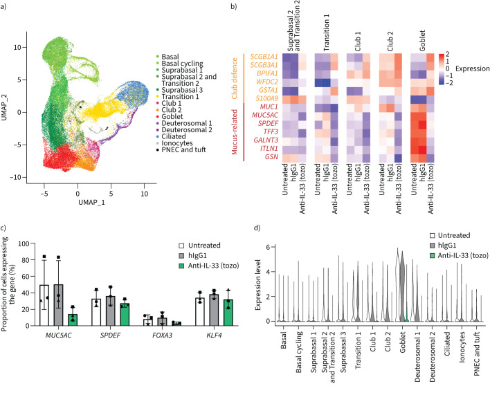
Methods: In vitro epithelial damage assays and three-dimensional, air-liquid interface (ALI) cell culture models of healthy and COPD epithelia were used to elucidate the functional role of IL-33ox. Transcriptomic changes occurring in healthy ALI cultures treated with IL-33ox and COPD ALI cultures treated with an IL-33-neutralising antibody were assessed with bulk and single-cell RNA sequencing analysis.

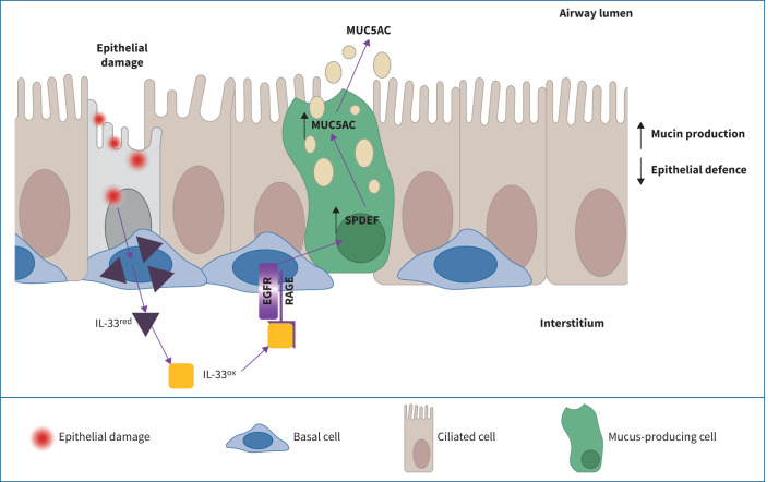
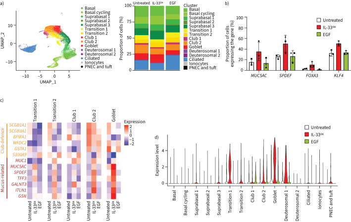
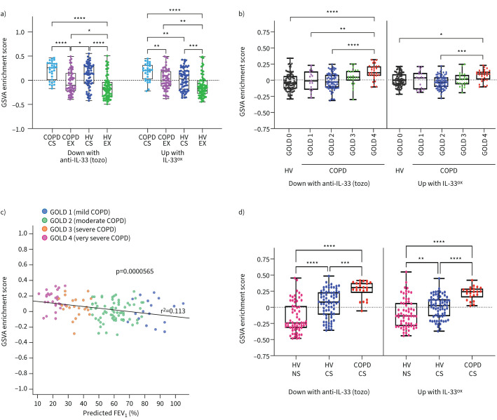
Results: We demonstrate that IL-33ox forms a complex with receptor for advanced glycation end products (RAGE) and epidermal growth factor receptor (EGFR) expressed on airway epithelium. Activation of this alternative, ST2-independent pathway impaired epithelial wound closure and induced airway epithelial remodelling in vitro. IL-33ox increased the proportion of mucus-producing cells and reduced epithelial defence functions, mimicking pathogenic traits of COPD. Neutralisation of the IL-33ox pathway reversed these deleterious traits in COPD epithelia. Gene signatures defining the pathogenic effects of IL-33ox were enriched in airway epithelia from patients with severe COPD.

Conclusions: Our study reveals for the first time that IL-33, RAGE and EGFR act together in an ST2-independent pathway in the airway epithelium and govern abnormal epithelial remodelling and muco-obstructive features in COPD.


Conflict of interest statement

Conflict of interest: S. Strickson, V.A. Negri, Y. Ohne, T. Ottosson, R.B. Dodd, C.C. Huntington, T. Baker, J. Li, K.E. Stephenson, A.J. O'Connor, J.S. Sagawe, H. Killick, D.G. Rees, S. Koch, Y. Wang, M. Ghaedi, S. Saumyaa, G.P. Sims, I.C. Scott, X. Romero Ros and E.S. Cohen are employees of AstraZeneca and may hold stock or stock options in AstraZeneca. K.F. Houslay, T. Moore, E. Gubbins, R. Kolbeck and A.A. Humbles are former employees of AstraZeneca and may hold stock or stock options in AstraZeneca. C. Sanden has nothing to disclose. J.S. Erjefält is a founder and board member of Medetect AB.

Figures

None
IL-33ox binds to receptor for advanced glycation end products (RAGE) to signal via epidermal growth factor receptor (EGFR). Activation of the IL-33ox–RAGE/EGFR pathway redirects epithelial cell fate, promoting a mucin hypersecretion phenotype at the expense of epithelial defence functions.
FIGURE 1
FIGURE 1
Oxidised interleukin 33 (IL-33ox) promotes the activation of epidermal growth factor receptor (EGFR). a) Representative images of scratch wound closure in submerged cultures at 0 h and 24 h shown on left (scale bar: 300 μm) and plotted as percentage wound closure at 24 h (middle) in growth factor-starved submerged normal human bronchial epithelial (NHBE) cells following treatment with serum stimulation-2 (ST2)-neutralising antibody (goat), IL-33-neutralising antibody (tozorakimab (tozo)) or human (hIgG1) or goat (gIgG1) immunoglobulin G1 isotype control antibody versus untreated control (five individual donors). Right, scratch wound closure in submerged NHBE cells at 24 h following treatment with reduced IL-33 (IL-33red), oxidation-resistant mutant IL-33 (IL-33C>S), IL-33ox, mouse immunoglobulin G1 (mIgG1) isotype control antibody, ST2-neutralising antibody, IL-33ox+mIgG1 isotype control antibody or IL-33ox+ST2-neutralising antibody versus untreated control (four individual donors). b) Phospho-proteome profiler MAPK signalling array analysis in submerged NHBE cells treated with 30 ng·mL−1 of IL-33ox, IL-33red or IL-33C>S for 10 min (one donor). c) Phospho-tyrosine kinase receptor array analysis in growth factor-starved submerged NHBE cells treated with 30 ng·mL−1 of IL-33red, IL-33C>S or IL-33ox for 10 min versus untreated control (one donor). p-EGFR: phosphorylated epidermal growth factor receptor. d) Western blot analysis of p-EGFR, total EGFR, p-STAT5, p-ERK1/2, p-JNK1/2 and p-p38 MAPK over time in submerged NHBE cells following stimulation with 30 ng·mL−1 of epidermal growth factor (EGF) or IL-33ox. e) Scratch wound closure in growth factor-starved submerged NHBE cells at 24 h following treatment with IL-33ox, IL-33ox+EGFR-neutralising antibody or IL-33ox+mIgG1 isotype control antibody versus untreated control (four individual donors in experimental replicate). The data shown are a representative example from at least two independent experiments. Individual data points are shown in panels a and e, and further details of box plots can be found in the supplementary materials; data are normalised to untreated control. *: p≤0.05; ****: p≤0.0001 (non-parametric Kruskal–Wallis test with multiple comparisons). ns: not significant.
FIGURE 2
FIGURE 2
Receptor for advanced glycation end products (RAGE) binds to oxidised interleukin 33 (IL-33ox), enabling the formation of a complex with epidermal growth factor receptor (EGFR). a) Left, schematic showing methodology for immunoprecipitation (IP) of EGFR in submerged normal human bronchial epithelial (NHBE) cells and tandem mass spectrometry (MS/MS). Right, quantitative Venn diagram showing the overlap of proteins detected by MS/MS following IP of EGFR in submerged NHBE cells treated with IL-33ox or epidermal growth factor (EGF) for 10 min. Proteins with a total unique peptide count of ≥3 in either treatment group are shown. Proteins overlapping with untreated control and IL-33ox are not shown. b) Western blot analysis of i) EGFR, RAGE and IL-33 following IP of EGFR in submerged NHBE cells treated with reduced IL-33 (IL-33red), IL-33ox or EGF for 10 min versus untreated control (one individual donor); ii) EGFR, RAGE and IL-33 following IP of EGFR in A549 cells or RAGE knockout (KO) A549 cells treated with IL-33ox for 0, 5, 10 or 15 min. c) Direct ELISA detecting binding of RAGE-Fc to immobilised IL-33red, oxidation-resistant mutant IL-33 (IL-33C>S), IL-33ox, high mobility group box 1 (HMGB1) or EGF. Absorption at 450 nm is shown. Error bars show sem. d) Scratch wound closure in submerged NHBE cells at 24 h following treatment with IL-33red, IL-33ox, IL-33ox+RAGE-neutralising antibody, IL-33ox+EGFR-neutralising antibody or IL-33ox+mouse immunoglobulin G1 (mIgG1) isotype control antibody versus untreated control (six individual donors). The data shown in b–d are representative examples from at least two independent experiments. Individual data points are shown, and further details of box plots can be found in the supplementary materials; data are normalised to untreated control. *: p≤0.05; **: p≤0.01 (non-parametric Kruskal–Wallis test with multiple comparisons).
FIGURE 3
FIGURE 3
Chronic oxidised interleukin 33 (IL-33ox) exposure induces an epithelial mucin hypersecretion phenotype. a) Schematic representation of bronchial epithelial air–liquid interface (ALI) cultures and end-point assays. FACS: fluorescence-activated cell sorting; IHC: immunohistochemistry; NHBE: normal human bronchial epithelial; scRNA seq: single-cell RNA sequencing. b) Heat map showing expression levels of genes in bronchial ALI cultures with significant variation between untreated control and oxidation-resistant mutant IL-33 (IL-33C>S), epidermal growth factor (EGF) and IL-33ox treatments from bulk RNA sequencing (three individual donors) (ANOVA, false discovery rate <0.0001). c) Representative IHC of healthy bronchial ALI cultures following a 7-day treatment with 30 ng·mL−1 of IL-33ox or EGF versus untreated control: mucin 5AC/B (MUC5AC/B) for goblet cells (yellow), acetylated α-tubulin for ciliated cells (teal) and p63 for basal cells (purple) (scale bar: 70 µm). d) Quantification of MUC5AC single-positive goblet cells as judged by flow cytometry in bronchial ALI cultures (six individual donors). e) ELISA of MUC5AC secreted into the apical region of healthy bronchial ALI cultures stimulated with IL-33C>S, IL-33ox or EGF versus untreated control (seven individual donors). **: p≤0.01 (non-parametric Kruskal–Wallis test with multiple comparisons).
FIGURE 4
FIGURE 4
Single-cell gene expression analysis identifies cell state changes supporting mucus hypersecretion. a) Left, UMAP plot of healthy bronchial epithelial air–liquid interface (ALI) cultures (three individual donors; 43 150 cells/single-cell transcriptomes) displaying the annotated cell states/types. Right, visual representation of changes in the proportions of cell states/types in bronchial ALI cultures following 7-day treatment with oxidised interleukin 33 (IL-33ox) or epidermal growth factor (EGF) (30 ng·mL−1) versus untreated control. b) Changes in the percentages of cells expressing mucin-related genes (MUC5AC, SPDEF, FOXA3 and KLF4) in bronchial ALI cultures after treatment with IL-33ox or EGF versus untreated control. Each symbol represents one healthy donor. Error bars show sem. c) Heat map showing the scale-normalised average expression levels of genes associated with mucin production or defence in the secretory states in bronchial ALI cultures treated with IL-33ox or EGF versus untreated control. d) Expression levels of MUC5AC in the annotated cell states/types in healthy bronchial ALI cultures treated with IL-33ox or EGF versus untreated control. PNEC: pulmonary neuroendocrine cell.
FIGURE 5
FIGURE 5
Inhibition of endogenous oxidised interleukin 33 (IL-33ox) reduces chronic obstructive pulmonary disease (COPD) epithelial dysfunction and mucus hypersecretion. a) Representative immunohistochemistry of COPD bronchial epithelial air–liquid interface (ALI) cultures following treatment with IL-33-neutralising antibody (tozorakimab (tozo)) or human immunoglobulin G1 (hIgG1) isotype control antibody (1 μg·mL−1) for 7 days: mucin 5AC/B (MUC5AC/B) for goblet cells (yellow), acetylated α-tubulin for ciliated cells (teal) and p63 for basal cells (purple) (scale bar: 70 µm). b) Flow cytometry analysis of intracellular MUC5AC in dissociated healthy or COPD bronchial ALI cultures following treatment with tozorakimab, serum stimulation-2 (ST2)-neutralising antibody or the relevant isotype control antibody versus untreated control (six individual healthy and five individual COPD donors). c) ELISA of MUC5AC secreted into the apical region of healthy or COPD ALI cultures incubated with tozorakimab, ST2-neutralising antibody or the relevant isotype control antibody versus untreated control (seven individual healthy and six individual COPD donors). d) MUC5AC expression determined by reverse transcription quantitative PCR in healthy or COPD bronchial ALI cultures incubated with tozorakimab, receptor for advanced glycation end products (RAGE)-neutralising antibody, endothelial growth factor receptor (EGFR)-neutralising antibody or the relevant isotype control antibody versus untreated control (four individual healthy and five individual COPD donors). Single data points from individual donors are shown; data are normalised to one donor in the untreated control group. e) Heat map showing expression levels of genes with significant variation between untreated control and tozorakimab, ST2-neutralising or relevant isotype control treatments from bulk RNA sequencing (three individual donors) (ANOVA, false discovery rate <0.0001). Individual data points are shown in panels b–d, and further details of box plots can be found in the supplementary materials. mIgG1: mouse immunoglobulin G1. *: p≤0.05; **: p≤0.01 (non-parametric Kruskal–Wallis test with multiple comparisons).
FIGURE 6
FIGURE 6
Inhibition of oxidised interleukin 33 (IL-33ox) reverses chronic obstructive pulmonary disease (COPD)-related cell state changes at the single-cell level. a) UMAP plot of COPD bronchial epithelial air–liquid interface (ALI) cultures treated with IL-33-neutralising antibody (tozorakimab (tozo)) or isotype control antibody versus untreated control (three individual donors; 60 137 cells from all conditions), showing the annotated cell states/types. b) Heat map showing the scale-normalised average expression levels of genes associated with mucin production or defence in the secretory states in COPD bronchial ALI cultures treated with tozorakimab or human immunoglobulin G1 (hIgG1) isotype control antibody versus untreated control. c) Changes in the percentages of cells expressing mucin-related genes (MUC5AC, SPDEF, FOXA3 and KLF4) in COPD bronchial ALI cultures after treatment with tozorakimab or hIgG1 isotype control antibody versus untreated control. Each symbol represents one COPD donor. Error bars show sem. d) MUC5AC expression levels in the annotated cell states/types in COPD bronchial ALI cultures treated with tozorakimab or hIgG1 isotype control antibody versus untreated control.
FIGURE 7
FIGURE 7
Gene signatures defining how the effects of oxidised interleukin 33 (IL-33ox) in bronchial epithelial air–liquid interface (ALI) cultures correlate with disease severity in patients with chronic obstructive pulmonary disease (COPD). a) Gene set variation analysis (GSVA) of the tozorakimab (tozo) and IL-33ox signatures in the GSE37147 cohort samples. b) GSVA showing the correlation with disease severity in the GSE47460 cohort samples derived from lung explants, as assessed by gene enrichment according to Global Initiative for Chronic Obstructive Lung Disease (GOLD) stages. c) GSVA showing the correlation of genes downregulated in COPD ALI cultures following IL-33-neutralising antibody (tozorakimab) treatment with disease severity (GOLD1–4) in the GSE47460 cohort samples, as assessed by gene enrichment score according to predicted forced expiratory volume in 1 s (FEV1). d) GSVA showing the correlation with smoking status and COPD in the GSE11784 cohort samples derived from lung explants. Data are plotted as mean±sem. Details of box plots can be found in the supplementary materials. CS: current smoker; EX: ex-smoker; HV: healthy volunteer; NS: non-smoker. *: p≤0.05; **: p≤0.01; ***: p≤0.001; ****: p≤0.0001 (one-way ANOVA with Tukey's honest significant difference test).

Comment in

References

    1. Carlier FM, de Fays C, Pilette C. Epithelial barrier dysfunction in chronic respiratory diseases. Front Physiol 2021; 12: 691227. doi:10.3389/fphys.2021.691227 - DOI - PMC - PubMed
    1. Hogg JC, Timens W. The pathology of chronic obstructive pulmonary disease. Annu Rev Pathol 2009; 4: 435–459. doi:10.1146/annurev.pathol.4.110807.092145 - DOI - PubMed
    1. Barnes PJ. COPD 2020: new directions needed. Am J Physiol Lung Cell Mol Physiol 2020; 319: L884–L886. doi:10.1152/ajplung.00473.2020 - DOI - PubMed
    1. Gohy S, Carlier FM, Fregimilicka C, et al. . Altered generation of ciliated cells in chronic obstructive pulmonary disease. Sci Rep 2019; 9: 17963. doi:10.1038/s41598-019-54292-x - DOI - PMC - PubMed
    1. Rabe KF, Celli BR, Wechsler ME, et al. . Safety and efficacy of itepekimab in patients with moderate-to-severe COPD: a genetic association study and randomised, double-blind, phase 2a trial. Lancet Respir Med 2021; 9: 1288–1298. doi:10.1016/S2213-2600(21)00167-3 - DOI - PubMed

Publication types

MeSH terms