

Somatostatin Receptor-PET/CT/MRI of Head and Neck Neuroendocrine Tumors
- PMID: 37442593
- PMCID: PMC10411831
- DOI: 10.3174/ajnr.A7934
Somatostatin Receptor-PET/CT/MRI of Head and Neck Neuroendocrine Tumors
Abstract
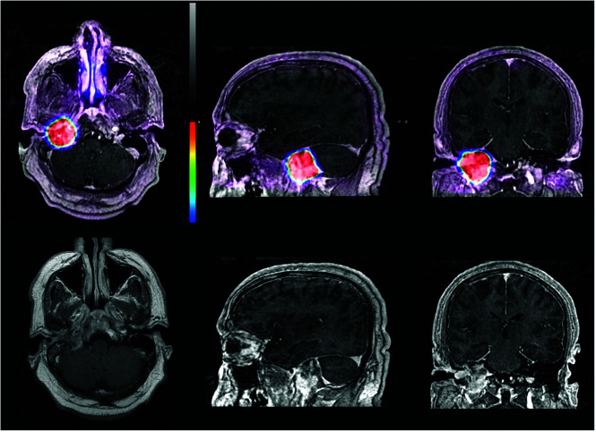
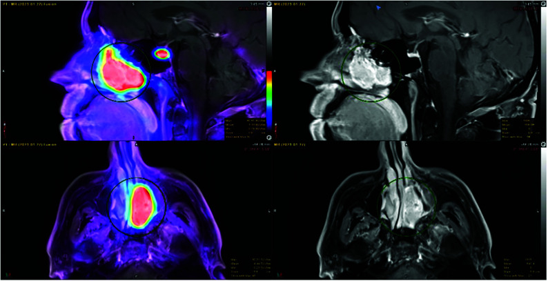
Background and purpose: Due to its high sensitivity, somatostatin receptor-PET may detect smaller lesions and more extensive disease than contrast-enhanced MR imaging, while the superior spatial resolution of MR imaging enables lesions to be accurately localized. We compared results of somatostatin receptor-PET/MRI with those of MR imaging alone and assessed the added value of vertex-to-thigh imaging for head and neck neuroendocrine tumors.
Materials and methods: Somatostatin receptor-PET/CT was acquired as limited brain or head and neck imaging, with optional vertex-to-thigh imaging, following administration of 64CU/68GA DOTATATE. Somatostatin receptor-PET was fused with separately acquired contrast-enhanced MR imaging. DOTATATE activity was classified as comparable, more extensive, and/or showing additional lesions compared with MR imaging. Vertex-to-thigh findings were classified as positive or negative for metastatic disease or incidental.
Results: Thirty patients (with 13 meningiomas, 11 paragangliomas, 1 metastatic papillary thyroid carcinoma, 1 middle ear neuroendocrine adenoma, 1 external auditory canal mass, 1 pituitary carcinoma, 1 olfactory neuroblastoma, 1 orbital mass) were imaged. Five had no evidence of somatostatin receptor-positive lesions and were excluded. In 11/25, somatostatin receptor-PET/MRI and MR imaging were comparable. In 7/25, somatostatin receptor-PET/MRI showed more extensive disease, while in 9/25, somatostatin receptor-PET/MRI identified additional lesions. On vertex-to-thigh imaging, 1 of 17 patients was positive for metastatic disease, 8 of 17 were negative, and 8 of 17 demonstrated incidental findings.
Conclusions: Somatostatin receptor-PET detected additional lesions and more extensive disease than contrast-enhanced MR imaging alone, while vertex-to-thigh imaging showed a low incidence of metastatic disease. Somatostatin receptor-PET/MRI enabled superior anatomic delineation of tumor burden, while any discrepancies were readily addressed. Somatostatin receptor-PET/MRI has the potential to play an important role in presurgical and radiation therapy planning of head and neck neuroendocrine tumors.
© 2023 by American Journal of Neuroradiology.
Figures


Similar articles
-
Hybrid Somatostatin Receptor PET/MRI of the Head and Neck.Radiographics. 2024 Oct;44(10):e240020. doi: 10.1148/rg.240020. Radiographics. 2024. PMID: 39325659 Review.
-
68Ga-DOTATATE PET/CT, 99mTc-HYNIC-octreotide SPECT/CT, and whole-body MR imaging in detection of neuroendocrine tumors: a prospective trial.J Nucl Med. 2014 Oct;55(10):1598-604. doi: 10.2967/jnumed.114.144543. Epub 2014 Aug 28. J Nucl Med. 2014. PMID: 25168627
-
The significance of incidental brain uptake on 68Ga-DOTATATE PET-CT in neuroendocrine tumour patients.Nucl Med Commun. 2016 Nov;37(11):1197-205. doi: 10.1097/MNM.0000000000000571. Nucl Med Commun. 2016. PMID: 27399848
-
Somatostatin receptor imaging in non-(131)I-avid metastatic differentiated thyroid carcinoma for determining the feasibility of peptide receptor radionuclide therapy with (177)Lu-DOTATATE: low fraction of patients suitable for peptide receptor radionuclide therapy and evidence of chromogranin A level-positive neuroendocrine differentiation.Clin Nucl Med. 2014 Jun;39(6):505-10. doi: 10.1097/RLU.0000000000000429. Clin Nucl Med. 2014. PMID: 24662668
-
Diagnostic value of [68 Ga]Ga-DOTA-labeled-somatostatin analogue PET/MRI for detecting liver metastasis in patients with neuroendocrine tumors: a systematic review and meta-analysis.Eur Radiol. 2022 Jul;32(7):4628-4637. doi: 10.1007/s00330-021-08527-z. Epub 2022 Jan 29. Eur Radiol. 2022. PMID: 35092473
Cited by
-
An Update on DOTA-Peptides PET Imaging and Potential Advancements of Radioligand Therapy in Intracranial Meningiomas.Life (Basel). 2025 Apr 7;15(4):617. doi: 10.3390/life15040617. Life (Basel). 2025. PMID: 40283171 Free PMC article. Review.
-
The Value of PET/CT in Particle Therapy Planning of Various Tumors with SSTR2 Receptor Expression: Comparative Interobserver Study.Cancers (Basel). 2024 May 15;16(10):1877. doi: 10.3390/cancers16101877. Cancers (Basel). 2024. PMID: 38791956 Free PMC article.
References
MeSH terms
Substances
LinkOut - more resources
Full Text Sources
Medical