. 2023 Nov;60(11):6275-6293.
doi: 10.1007/s12035-023-03444-2. Epub 2023 Jul 13.

Conservation of vCJD Strain Properties After Extraction and In Vitro Propagation of PrPSc from Archived Formalin-Fixed Brain and Appendix Tissues Using Highly Sensitive Protein Misfolding Cyclic Amplification

Affiliations

Conservation of vCJD Strain Properties After Extraction and In Vitro Propagation of PrPSc from Archived Formalin-Fixed Brain and Appendix Tissues Using Highly Sensitive Protein Misfolding Cyclic Amplification

Suzanne Suleiman et al. Mol Neurobiol. 2023 Nov.

Abstract

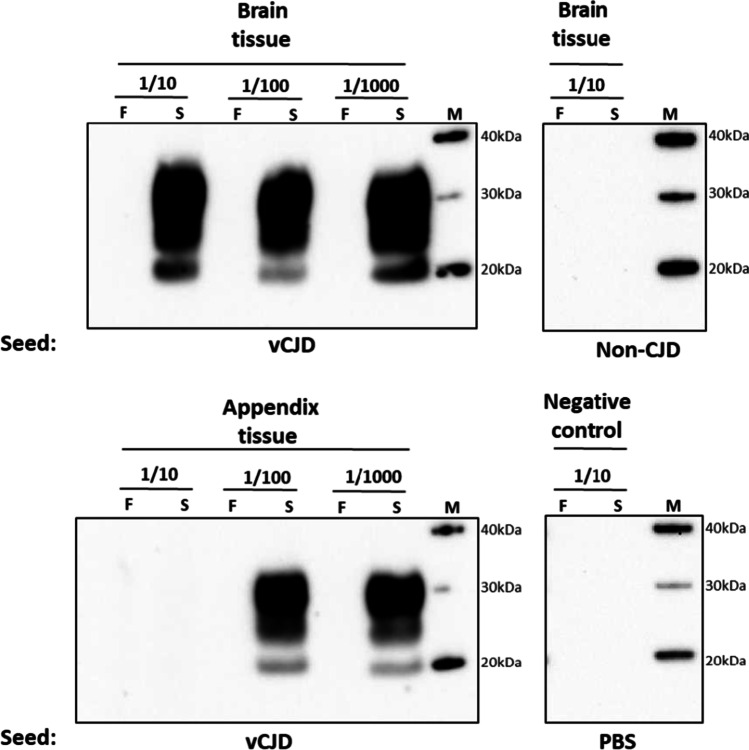
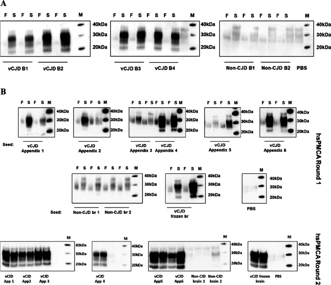
Three retrospective lymphoreticular tissue studies (Appendix I, II, and III) aimed to estimate the UK prevalence of variant Creutzfeldt-Jakob disease (vCJD), following exposure of the population to the bovine spongiform encephalopathy (BSE) agent, in the late 1980s and 1990s. These studies evaluated the presence of abnormal prion protein aggregates, in archived formalin-fixed paraffin-embedded (FFPE) appendectomy samples, by immunohistochemical detection. Although there was concordance in the estimated prevalence of vCJD from these studies, the identification of positive specimens from pre- and post-BSE-exposure periods in Appendix III study has raised questions regarding the nature and origin of the detected abnormal prion protein. We applied a robust and novel approach in the extraction of disease-associated prion protein (PrPSc) present in frozen and FFPE samples of brain and appendix from a patient with pathologically confirmed vCJD. The extracted material was used to seed the highly sensitive protein misfolding cyclic amplification assay (hsPMCA) to investigate the in vitro and in vivo propagation properties of the extracted abnormal prion protein. We demonstrate that PrPSc can be successfully extracted from FFPE appendix tissue and propagated in vitro. Bioassay in wild-type and gene-targeted mouse models confirmed that the extracted and amplified product is infectious and retains strain properties consistent with vCJD. This provides a highly sensitive and reliable platform for subsequent analysis of the archived FFPE appendix tissue derived from the Appendix II and III surveys, to further evaluate the nature of the abnormal PrP detected in the positive samples.

Keywords: Creutzfeldt-Jakob disease (CJD), Bovine spongiform encephalopathy (BSE); Neurodegenerative disorders; Prion; Protein misfolding; Protein misfolding cyclic amplification assay (PMCA).


Conflict of interest statement

The authors declare no competing interests.

Figures

Fig. 1
Fig. 1
Outline of the different preparations used for animal transmission experiments (inoculum I–VI). Frozen and FFPE brain and appendix tissues originated from a single case of vCJD were used in the preparation of all six inocula for inoculation in RIII and HuMM mice. Inoculum “I” corresponds to 10% tissue homogenate prepared by homogenising vCJD brain tissue in physiological saline. Inoculum “II” is similar to inoculum I, with the exception that the vCJD brain tissue material was homogenised in conversion buffer. Inoculum “III” corresponds to hsPMCA-amplified material prepared by serially propagating a 10% vCJD frozen brain homogenate in conversion buffer (dilution from 10% brain homogenate, corresponds to a dilution of 1 × 10−38). Inoculum “IV” corresponds to hsPMCA-amplified material prepared by serially amplifying a 10% frozen appendix homogenate in conversion buffer (dilution from 10% brain homogenate, corresponds to a dilution of 1 × 10−38). Inoculum “V” corresponds to hsPMCA-amplified material prepared by serially propagating a 10% extracted homogenate from FFPE vCJD brain in conversion buffer. Finally, inoculum “VI” corresponds to hsPMCA-amplified material prepared by serially propagating a 10% extracted homogenate from FFPE vCJD appendix in conversion buffer
Fig. 2
Fig. 2
Amplification of vCJD PrP.Sc by hsPMCA. Frozen brain and appendix tissues from a single vCJD case were homogenised and diluted 1:10, 1:100, and 1:1000 (from the 10% tissue homogenates) in larger volumes of codon 129MM transgenic mouse substrate for amplification. The reactions were subjected to a single round of hsPMCA following proteolytic treatment by PK and Western blot analysis using the 3F4 antibody. In addition to the PBS reaction, a brain tissue sample from a non-CJD case was used as a negative control. For comparison purposes, a small aliquot (19 µL) of each hsPMCA reaction was taken prior to the amplification step and stored at minus 80 °C (frozen sample “F” sample). The corresponding samples were harvested post-amplification (sonicated sample “S”), and both samples were tested for PrPres levels by Western blot. Western blot protein standard was included in each individual panel (M)
Fig. 3
Fig. 3
Protocol optimisation using vCJD FFPE human tissues. A Overview of the experimental strategy for retrieving PrPSc from FFPE tissues. The protocol was developed by testing four combinations comprising two deparaffinisation steps and two homogenisation methods. Deparaffinisation was performed either using xylene followed by treatment with 100%, 90%, and 70% grades of ethanol and a final wash with PBS (“Xy/Eth” method) or by three washes with water heated to 85 °C and PBS (“85 °C H2O” method). After centrifugation at 14,000 g for 10 min, the residual tissues are either homogenised by three cycles (45 s, 6 ms−1) of the FastPrep®-24 homogeniser (FP), or by manual homogenisation using a sterile plastic pestle (PP). B hsPMCA to assess the effectiveness and sensitivity of the four procedures using vCJD FFPE human brain sections. The ability of the different procedures to recover PrPSc aggregates from FFPE brain tissues was revealed by Western blot analysis of the hsPMCA amplified products (sonicated sample “S”) compared with the non-sonicated counterpart (“F” samples). One case of vCJD was used, and a serial dilution of 10−1–10−6 of the homogenised material was tested for each combination of procedures. C Effectiveness of the PrP.Sc extraction method in FFPE human appendix tissues. Amplification efficacy of the appendix material from a single definite vCJD case extracted and homogenised by (“85 °C H2O” + PP) procedure was evaluated with 1:10 and 1:100 seed: substrate ratios after a single (first panel) and a double (second panel) rounds of hsPMCA. In the first round of hsPMCA, the non-sonicated samples “F” were tested alongside sonicated samples “S”. All samples were assessed in duplicate, with at least three technical replica were performed for each sample. Positive control of amplified frozen vCJD brain homogenate was included in parallel to evaluate amplification efficacy. A negative control reaction, subjected to the amplification process, was also tested. Phosphate buffered saline “PBS”. Western blot protein standard (M)
Fig. 3
Fig. 3
Protocol optimisation using vCJD FFPE human tissues. A Overview of the experimental strategy for retrieving PrPSc from FFPE tissues. The protocol was developed by testing four combinations comprising two deparaffinisation steps and two homogenisation methods. Deparaffinisation was performed either using xylene followed by treatment with 100%, 90%, and 70% grades of ethanol and a final wash with PBS (“Xy/Eth” method) or by three washes with water heated to 85 °C and PBS (“85 °C H2O” method). After centrifugation at 14,000 g for 10 min, the residual tissues are either homogenised by three cycles (45 s, 6 ms−1) of the FastPrep®-24 homogeniser (FP), or by manual homogenisation using a sterile plastic pestle (PP). B hsPMCA to assess the effectiveness and sensitivity of the four procedures using vCJD FFPE human brain sections. The ability of the different procedures to recover PrPSc aggregates from FFPE brain tissues was revealed by Western blot analysis of the hsPMCA amplified products (sonicated sample “S”) compared with the non-sonicated counterpart (“F” samples). One case of vCJD was used, and a serial dilution of 10−1–10−6 of the homogenised material was tested for each combination of procedures. C Effectiveness of the PrP.Sc extraction method in FFPE human appendix tissues. Amplification efficacy of the appendix material from a single definite vCJD case extracted and homogenised by (“85 °C H2O” + PP) procedure was evaluated with 1:10 and 1:100 seed: substrate ratios after a single (first panel) and a double (second panel) rounds of hsPMCA. In the first round of hsPMCA, the non-sonicated samples “F” were tested alongside sonicated samples “S”. All samples were assessed in duplicate, with at least three technical replica were performed for each sample. Positive control of amplified frozen vCJD brain homogenate was included in parallel to evaluate amplification efficacy. A negative control reaction, subjected to the amplification process, was also tested. Phosphate buffered saline “PBS”. Western blot protein standard (M)
Fig. 4
Fig. 4
TSE vacuolation, vacuolation profiles, and PrPSc deposition in wild-type mice (RIII) and gene-targeted (HuMM) mice models challenged with frozen and FFPE brain and appendix tissue propagated by hsPMCA. TSE associated neuropathology in RIII and HuMM mice challenged with six vCJD inocula preparations (I) 10% vCJD brain homogenate in saline (circle), (II) 10% vCJD brain homogenate in PMCA conversion buffer (square), (III) hsPMCA vCJD brain product 1 × 10−38 (empty triangle), (IV) hsPMCA vCJD appendix product 1 × 10−38 (diamond), (V) hsPMCA vCJD FFPE brain product 1 × 10−38 (segmented line, black square), and (VI) hsPMCA vCJD FFPE appendix product 1 × 10.−38 (segmented line, empty circle). A TSE associated vacuolation in the medulla in RIII mice and the thalamus in RIII and HuMM mice. Arrow highlights amyloid plaque in the thalamus of the HuMM. B TSE vacuolation profiles in RIII and HuMM mice, n > 6 in each cohort. Profiles contain both clinical and non-clinical mice with TSE vacuolation. All data shows mean ± SEM. Brain region areas: G1–9, grey matter scoring areas; G1, medulla; G2, cerebellum; G3, superior colliculus; G4, hypothalamus; G5, thalamus; G6, hippocampus; G7, septum; G8, retrosplenial and adjacent motor cortex; G9, cingulate and adjacent motor cortex. W1–W3, white matter scoring regions: W1, cerebellar white matter; W2, mesencephalic tegmentum; W3, cerebral peduncle. C Abnormal PrP deposition in the hippocampus (inset of CA2 region) of RIII mice and thalamus of HuMM mice. RIII mice; scale bars = 200 µm, inset = 100 µm. Antibody: SAF83. HuMM mice; scale bars = 200 µm. Antibody: 3F4
Fig. 4
Fig. 4
TSE vacuolation, vacuolation profiles, and PrPSc deposition in wild-type mice (RIII) and gene-targeted (HuMM) mice models challenged with frozen and FFPE brain and appendix tissue propagated by hsPMCA. TSE associated neuropathology in RIII and HuMM mice challenged with six vCJD inocula preparations (I) 10% vCJD brain homogenate in saline (circle), (II) 10% vCJD brain homogenate in PMCA conversion buffer (square), (III) hsPMCA vCJD brain product 1 × 10−38 (empty triangle), (IV) hsPMCA vCJD appendix product 1 × 10−38 (diamond), (V) hsPMCA vCJD FFPE brain product 1 × 10−38 (segmented line, black square), and (VI) hsPMCA vCJD FFPE appendix product 1 × 10.−38 (segmented line, empty circle). A TSE associated vacuolation in the medulla in RIII mice and the thalamus in RIII and HuMM mice. Arrow highlights amyloid plaque in the thalamus of the HuMM. B TSE vacuolation profiles in RIII and HuMM mice, n > 6 in each cohort. Profiles contain both clinical and non-clinical mice with TSE vacuolation. All data shows mean ± SEM. Brain region areas: G1–9, grey matter scoring areas; G1, medulla; G2, cerebellum; G3, superior colliculus; G4, hypothalamus; G5, thalamus; G6, hippocampus; G7, septum; G8, retrosplenial and adjacent motor cortex; G9, cingulate and adjacent motor cortex. W1–W3, white matter scoring regions: W1, cerebellar white matter; W2, mesencephalic tegmentum; W3, cerebral peduncle. C Abnormal PrP deposition in the hippocampus (inset of CA2 region) of RIII mice and thalamus of HuMM mice. RIII mice; scale bars = 200 µm, inset = 100 µm. Antibody: SAF83. HuMM mice; scale bars = 200 µm. Antibody: 3F4
Fig. 5
Fig. 5
Western blot and PrPres detection in RIII and HuMM mice challenged with the four frozen (A) and two FFPE (B) inocula preparations. Western blots were performed to evaluate the presence of PrPres in frozen RIII and HuMM brains. Brain tissue was homogenised to prepare a 10% tissue homogenate in conversion buffer. After PK treatment, Western blot analysis was performed as described in the Western blot analysis section. A Inoculum “I”, 10% vCJD brain homogenate in saline; inoculum “II”, 10% vCJD brain homogenate in PMCA conversion buffer; inoculum “III”, hsPMCA vCJD brain product 1 × 10−38; and inoculum “IV”, hsPMCA vCJD appendix product 1 × 10−38. B Inoculum “V” hsPMCA vCJD FFPE brain product 1 × 10−38; and inoculum “VI”, hsPMCA vCJD FFPE appendix product 1 × 10−38. On each gel, a type “2B” vCJD reference sample was used as a positive control (minus and plus PK treatment). Monoclonal antibody 6H4 used for RIII. Monoclonal antibody 3F4 used for HuMM mice. (*): indicates where NaPTA precipitation step was perfumed to concentrate PrP.Sc in HuMM brain homogenate samples. PK, proteinase K. NaPTA, sodium phosphotungstate. Western blot protein standard (M)
Fig. 6
Fig. 6
Protocol evaluation for extraction and amplification of the vCJD PrP.Sc from FFPE human brain and appendix tissues. A Evaluation of the selected protocol using FFPE human brain tissue sections of different vCJD and non-CJD cases. FFPE human brain tissue sections of four vCJD and two non-CJD cases were deparaffinised by 85 °C heated water and homogenised by sterile plastic pestle. The extracted material was used to seed an hsPMCA reaction in a 1:1000 seed: substrate ratio. The reactions where then analysed by PK-digestion and western blotting. All cases were run in duplicate and the amplification of the sonicated samples “S” was compared to the non-sonicated mix “F”. A blank reaction was performed by seeding the same substrate with PBS. B Protocol evaluation using FFPE human appendix tissues from different vCJD cases. FFPE human appendix tissue from six definite vCJD cases and FFPE brain tissue from two non-CJD brain cases, processed by the same protocol as above, were subjected to single (first panel) and doubled (second panel) rounds of hsPMCA in a 1:10 seed: substrate ratio. The reactions were analysed by PK-digestion and western blotting. In parallel, one vCJD brain homogenate was included as a positive control. Unseeded reaction included with PBS was used as a blank reaction. In the first round of hsPMCA, sonicated samples “S” were compared with their non-sonicated counterparts “F”. All samples were run in duplicates in the consecutive rounds of hsPMCA. Western blot protein standard “M”

Similar articles

References

    1. Will RG, Ironside JW, Zeidler M, Cousens SN, Estibeiro K, Alperovitch A, Poser S, Pocchiari M, Hofman A, Smith PG. A new variant of Creutzfeldt-Jakob disease in the UK. Lancet. 1996;347(9006):921–925. doi: 10.1016/S0140-6736(96)91412-9. - DOI - PubMed
    1. Collinge J, Sidle KC, Meads J, Ironside J, Hill AF. Molecular analysis of prion strain variation and the aetiology of ‘new variant’ CJD. Nature. 1996;383(6602):685–690. doi: 10.1038/383685a0. - DOI - PubMed
    1. Parchi P, Capellari S, Chen SG, Petersen RB, Gambetti P, Kopp N, Brown P, Kitamoto T, Tateishi J, Giese A, Kretzschmar H. Typing prion isoforms. Nature. 1997;386(6622):232–234. doi: 10.1038/386232a0. - DOI - PubMed
    1. Hill AF, Zeidler M, Ironside J, Collinge J. Diagnosis of new variant Creutzfeldt-Jakob disease by tonsil biopsy. Lancet. 1997;349(9045):99–100. doi: 10.1016/S0140-6736(97)24002-X. - DOI - PubMed
    1. Hilton DA, Fathers E, Edwards P, Ironside JW, Zajicek J. Prion immunoreactivity in appendix before clinical onset of variant Creutzfeldt-Jakob disease. Lancet. 1998;352(9129):703–704. doi: 10.1016/S0140-6736(98)24035-9. - DOI - PubMed