

Glycogen availability and pH variation in a medium simulating vaginal fluid influence the growth of vaginal Lactobacillus species and Gardnerella vaginalis
- PMID: 37442975
- PMCID: PMC10339506
- DOI: 10.1186/s12866-023-02916-8
Glycogen availability and pH variation in a medium simulating vaginal fluid influence the growth of vaginal Lactobacillus species and Gardnerella vaginalis
Abstract
Background: Glycogen metabolism by Lactobacillus spp. that dominate the healthy vaginal microbiome contributes to a low vaginal pH (3.5-4.5). During bacterial vaginosis (BV), strict and facultative anaerobes including Gardnerella vaginalis become predominant, leading to an increase in the vaginal pH (> 4.5). BV enhances the risk of obstetrical complications, acquisition of sexually transmitted infections, and cervical cancer. Factors critical for the maintenance of the healthy vaginal microbiome or the transition to the BV microbiome are not well defined. Vaginal pH may affect glycogen metabolism by the vaginal microflora, thus influencing the shift in the vaginal microbiome.
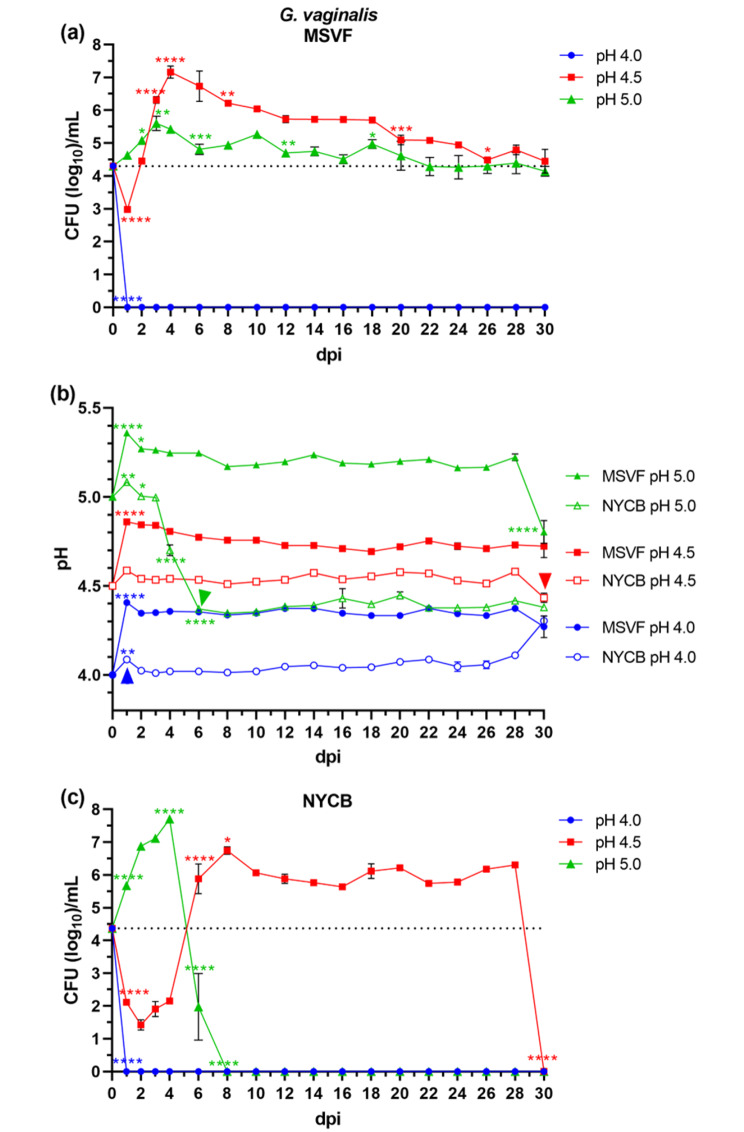
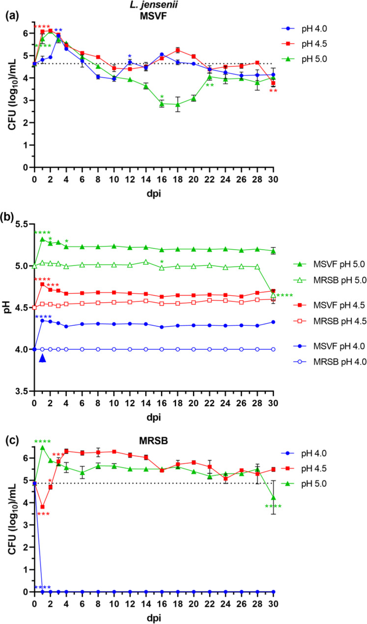
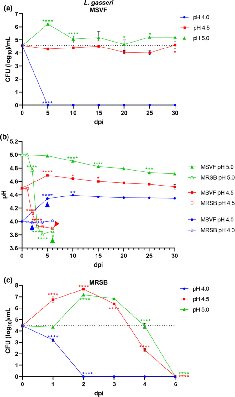
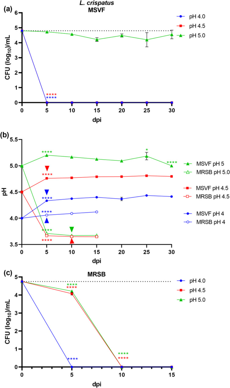
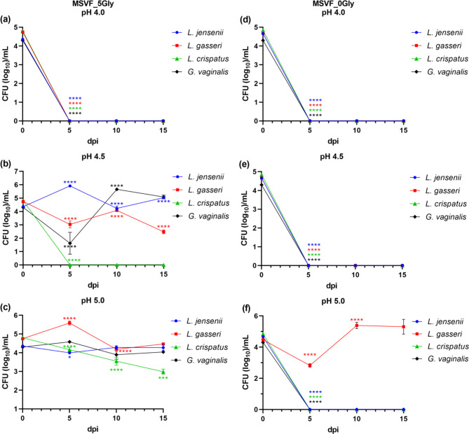
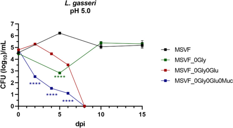
Results: The medium simulating vaginal fluid (MSVF) supported growth of L. jensenii 62G, L. gasseri 63 AM, and L. crispatus JV-V01, and G. vaginalis JCP8151A at specific initial pH conditions for 30 d. L. jensenii at all three starting pH levels (pH 4.0, 4.5, and 5.0), G. vaginalis at pH 4.5 and 5.0, and L. gasseri at pH 5.0 exhibited the long-term stationary phase when grown in MSVF. L. gasseri at pH 4.5 and L. crispatus at pH 5.0 displayed an extended lag phase over 30 d suggesting inefficient glycogen metabolism. Glycogen was essential for the growth of L. jensenii, L. crispatus, and G. vaginalis; only L. gasseri was able to survive in MSVF without glycogen, and only at pH 5.0, where it used glucose. All four species were able to survive for 15 d in MSVF with half the glycogen content but only at specific starting pH levels - pH 4.5 and 5.0 for L. jensenii, L. gasseri, and G. vaginalis and pH 5.0 for L. crispatus.
Conclusions: These results suggest that variations in the vaginal pH critically influence the colonization of the vaginal tract by lactobacilli and G. vaginalis JCP8151A by affecting their ability to metabolize glycogen. Further, we found that L. jensenii 62G is capable of glycogen metabolism over a broader pH range (4.0-5.0) while L. crispatus JV-V01 glycogen utilization is pH sensitive (only functional at pH 5.0). Finally, our results showed that G. vaginalis JCP8151A can colonize the vaginal tract for an extended period as long as the pH remains at 4.5 or above.
Keywords: Gardnerella vaginalis; Glucose; Glycogen; Lactobacillus crispatus; Lactobacillus gasseri; Lactobacillus jensenii; Medium simulating vaginal fluid; pH.
© 2023. The Author(s).
Conflict of interest statement
The authors declare that they have no competing interests.
Figures
Similar articles
-
Comparison of main lactobacillus species between healthy women and women with bacterial vaginosis.Chin Med J (Engl). 2009 Nov 20;122(22):2748-51. Chin Med J (Engl). 2009. PMID: 19951608
-
Influence of Lactobacillus crispatus, Lactobacillus iners and Gardnerella vaginalis on bacterial vaginal composition in pregnant women.Arch Gynecol Obstet. 2021 Aug;304(2):395-400. doi: 10.1007/s00404-021-05978-z. Epub 2021 Feb 1. Arch Gynecol Obstet. 2021. PMID: 33521838
-
Quantitative determination by real-time PCR of four vaginal Lactobacillus species, Gardnerella vaginalis and Atopobium vaginae indicates an inverse relationship between L. gasseri and L. iners.BMC Microbiol. 2007 Dec 19;7:115. doi: 10.1186/1471-2180-7-115. BMC Microbiol. 2007. PMID: 18093311 Free PMC article.
-
[Characteristics and physiologic role of female lower genital microbiome].Orv Hetil. 2023 Jun 18;164(24):923-930. doi: 10.1556/650.2023.32791. Print 2023 Jun 18. Orv Hetil. 2023. PMID: 37330978 Review. Hungarian.
-
Vaginal microbiome.Ceska Gynekol. 2018 Winter;83(5):371-379. Ceska Gynekol. 2018. PMID: 30848142 Review. English.
Cited by
-
Dual probiotic and antibiotic therapy targeting bacterial vaginosis: an integrated experimental/computational modeling perspective.Biomed Eng Adv. 2025 Jun;9:100163. doi: 10.1016/j.bea.2025.100163. Epub 2025 Apr 2. Biomed Eng Adv. 2025. PMID: 40529156
-
Identification and characterisation of vaginal bacteria-glycan interactions implicated in reproductive tract health and pregnancy outcomes.Nat Commun. 2025 Jun 5;16(1):5207. doi: 10.1038/s41467-025-60404-1. Nat Commun. 2025. PMID: 40467588 Free PMC article.
-
Leveraging the microbiome to combat antibiotic resistant gynecological infections.NPJ Antimicrob Resist. 2025 Apr 23;3(1):32. doi: 10.1038/s44259-025-00106-2. NPJ Antimicrob Resist. 2025. PMID: 40269132 Free PMC article. Review.
-
Navigating the Vaginal Milieu During Perimenopause: A Narrative Review of Physiological Changes and Clinical Implications.J Pharm Bioallied Sci. 2025 May;17(Suppl 1):S92-S95. doi: 10.4103/jpbs.jpbs_1493_24. Epub 2025 Apr 21. J Pharm Bioallied Sci. 2025. PMID: 40511019 Free PMC article. Review.
-
Genital cutaneous candidiasis versus chronic recurrent vulvovaginal candidiasis: distinct diseases, different populations.Clin Microbiol Rev. 2025 Jun 12;38(2):e0002025. doi: 10.1128/cmr.00020-25. Epub 2025 May 28. Clin Microbiol Rev. 2025. PMID: 40434101 Review.
References
-
- Aldunate M, Srbinovski D, Hearps AC, Latham CF, Ramsland PA, Gugasyan R, et al. Antimicrobial and immune modulatory effects of lactic acid and short chain fatty acids produced by vaginal microbiota associated with eubiosis and bacterial vaginosis. Front Physiol. 2015;6:164. doi: 10.3389/fphys.2015.00164. - DOI - PMC - PubMed
-
- Witkin SS, Linhares IM. Why do lactobacilli dominate the human vaginal microbiota? BJOG. 2017;124(4):606–11; 10.1111/1471-0528.14390. - PubMed
Publication types
MeSH terms
Substances
LinkOut - more resources
Full Text Sources
Miscellaneous
