. 2023 Jul 13;21(1):158.
doi: 10.1186/s12915-023-01655-6.

Identification of residues critical for the extension of Munc18-1 domain 3a

Affiliations

Identification of residues critical for the extension of Munc18-1 domain 3a

Xianping Wang et al. BMC Biol. .

Abstract

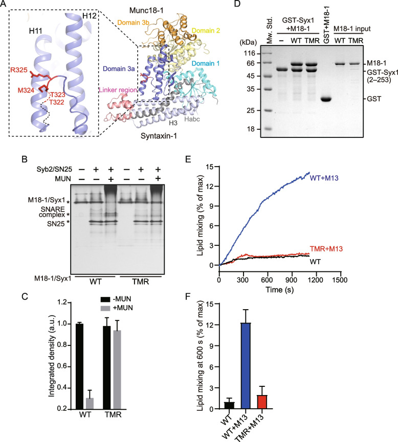
Background: Neurotransmitter release depends on the fusion of synaptic vesicles with the presynaptic membrane and is mainly mediated by SNARE complex assembly. During the transition of Munc18-1/Syntaxin-1 to the SNARE complex, the opening of the Syntaxin-1 linker region catalyzed by Munc13-1 leads to the extension of the domain 3a hinge loop, which enables domain 3a to bind SNARE motifs in Synaptobrevin-2 and Syntaxin-1 and template the SNARE complex assembly. However, the exact mechanism of domain 3a extension remains elusive.

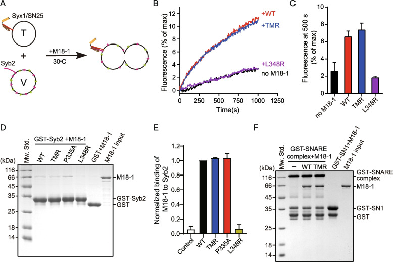
Results: Here, we characterized residues on the domain 3a hinge loop that are crucial for the extension of domain 3a by using biophysical and biochemical approaches and electrophysiological recordings. We showed that the mutation of residues T323/M324/R325 disrupted Munc13-1-mediated SNARE complex assembly and membrane fusion starting from Munc18-1/Syntaxin-1 in vitro and caused severe defects in the synaptic exocytosis of mouse cortex neurons in vivo. Moreover, the mutation had no effect on the binding of Synaptobrevin-2 to isolated Munc18-1 or the conformational change of the Syntaxin-1 linker region catalyzed by the Munc13-1 MUN domain. However, the extension of the domain 3a hinge loop in Munc18-1/Syntaxin-1 was completely disrupted by the mutation, leading to the failure of Synaptobrevin-2 binding to Munc18-1/Syntaxin-1.

Conclusions: Together with previous results, our data further support the model that the template function of Munc18-1 in SNARE complex assembly requires the extension of domain 3a, and particular residues in the domain 3a hinge loop are crucial for the autoinhibitory release of domain 3a after the MUN domain opens the Syntaxin-1 linker region.

Keywords: Conformational change; Munc13-1; Munc18-1; SNARE complex; Synaptic exocytosis; Syntaxin-1.


Conflict of interest statement

The authors declare that they have no competing interests.

Figures

Fig. 1
Fig. 1
Residues T323/M324/R325 are essential for SNARE complex assembly from Munc18-1/Syx1 catalyzed by the MUN domain. A Crystal structure of Munc18-1/Syx1 (PDB ID: 3C98) and positions indicating mutation sites (T322, T323, M324 and R325). Mutation sites are highlighted in red, and the residue sequence (317–323) without electron density is shown as dashed lines. B The triple mutation T323A/M324A/R325A (TMR) completely abolished the transition from Munc18-1/Syx1 to the SNARE complex catalyzed by the MUN domain, as shown by native PAGE. C Quantification of the results shown in B. Data are presented as mean values ± SD; n = 3. D The TMR mutation had no effect on the interaction between Munc18-1 and Syx1, as detected by GST pull-down assay. E Effect of TMR mutation on fusion between liposomes containing Munc18-1/Syx1 and liposomes reconstituted with Syb2 in the presence of SNAP-25, the Syt1 C2AB fragment, and 1 mM CaCl2, with or without the Munc13-1 C1C2BMUN fragment. F The quantification results of data shown in E. Data are presented as mean values ± SD; n = 3. Representative gel displayed is from one of three independent replicates. M18-1, Munc18-1; Syx1, Syntaxin-1; M13, Munc13-1 C1C2BMUN domain; SN25, SNAP-25; Syb2, Synaptobrevin-2
Fig. 2
Fig. 2
Residues T323/M324/R325 are crucial for synaptic vesicle exocytosis. A Sample traces (left), summary graphs of frequency (middle), and amplitude (right) of mIPSC. Data were recorded from cultured cortical neurons infected with control lentivirus (control), lentivirus expressing Munc18-1 shRNA (none), and rescued expressing sequences of Munc18-1 WT (WT) or Munc18-1 TMR mutant (TMR). B Sample traces (left), summary graphs of amplitude (middle), and charge transfer (right) of evoked IPSCs triggered by action potential recorded from cultured cortical neurons as described in A. C Sample traces (left) and summary graphs of charge transfer (right) of hypertonic sucrose-evoked IPSC recorded from cultured cortical neurons as described in A. Data are presented as mean ± SEM. Statistical significance was analyzed by Student’s t test; **, P < 0.01; ***, P < 0.001. Cells recorded are from at least three independent cultures, and the cell numbers are shown in bars. KD, knockdown
Fig. 3
Fig. 3
The TMR mutation supports Syx1 transport. A Representative images of HEK293T cells transfected with plasmids expressing EGFP-Munc18-1 and mCherry2-Syx1 respectively. B Representative images of HEK293T cells cotransfected with mCherry2-Syx1 and EGFP-Munc18-1 WT (WT) or TMR mutant (TMR). C Quantification of Munc18-1 and Syx1 distribution on the plasma membrane. Pictures used for analysis are from three independent cultures, and the numbers are listed in the bars. Data are shown as mean values ± SEM. Statistical significance was analyzed by Student’s t test. ***, P < 0.001. Scale bar: 20 μm; PM: plasma membrane
Fig. 4
Fig. 4
The TMR mutation supported Syb2 binding to isolated Munc18-1. A Illustration of Munc18-1-promoted reconstituted liposome fusion between v-liposomes bearing Syb2 (1–116) and t-liposomes containing Syx1 (1–288)/SNAP-25. V-liposomes were prepared by including NBD-PE and Rhodamine-PE, and NBD fluorescence at 538 nm was monitored. B Traces of lipid mixing reactions promoted by Munc18-1. Traces represented are from one of three independent replicates. All reactions were performed at 30 °C. C Quantification of the results shown in B. Data are presented as mean ± SD, n = 3. D Interaction between Munc18-1 and Syb2 detected by GST pull-down assay. The representative gel shown was from one of three replicates. E Quantification of integrated densities of Munc18-1 bands. The band densityy of WT Munc18-1 was taken as 1. Data are presented as mean values ± SD, n = 3. F Effect of TMR mutation on the Munc18-1 binding SNARE complex detected by GST pull-down assay. The SNARE complex was assembled by incubating GST-SN1, SN3, the cytoplasmic domain of Syb2 (29–93) and Syx1 (2–253) overnight at 4 °C. Bands of Munc18-1 and the SNARE complex are indicated in the figure. Representative gel displayed is from one of three independent replicates
Fig. 5
Fig. 5
Crucial role of residues T323/M324/R325 in the extension of domain 3a hinge loop. A Position of FRET pair labeled on Syx1 to monitor the relative movement between the linker region and Habc in the crystal structure of Munc18-1/Syx1 (PDB ID: 3C98). A Syx1 S95C/C145S/S171C mutation was generated for stochastic labeling of the FRET pair Alexa-555 and Alexa-647. B Representative histograms of smFRET efficiency of FRET-pairs labeled Syx1 binding Munc18-1 WT or TMR mutant in the absence and presence of MUN domain. Munc18-1/Syx1 was fixed on a coverslip modified with PEG/PEG-NTA-Co2+ via the His tag at the C-terminus of Syx1. C TMR mutation impaired Syb2 binding to the Munc18-1/Syx1 complex in the presence of the MUN domain, as detected by GST pull-down. Bands of Munc18-1 were highlighted with red asterisk in Coomassie brilliant blue stained SDS–PAGE. Representative gel displayed is from one of three independent replicates. D Quantification of the integrated densities of the Munc18-1 bands in C. The band density of WT Munc18-1 in the presence of the MUN domain was taken as 1. Data are presented as mean values ± SD, n = 3

Similar articles

Cited by

References

    1. Südhof TC. Calcium control of neurotransmitter release. Cold Spring Harb Perspect Biol. 2012;4(1):a011353. doi: 10.1101/cshperspect.a011353. - DOI - PMC - PubMed
    1. Jahn R, Fasshauer D. Molecular machines governing exocytosis of synaptic vesicles. Nature. 2012;490(7419):201–207. doi: 10.1038/nature11320. - DOI - PMC - PubMed
    1. Südhof TC. Neurotransmitter release: the last millisecond in the life of a synaptic vesicle. Neuron. 2013;80(3):675–690. doi: 10.1016/j.neuron.2013.10.022. - DOI - PMC - PubMed
    1. Südhof TC, Rizo J. Synaptic vesicle exocytosis. Cold Spring Harb Perspect Biol. 2011;3(12):a005637. doi: 10.1101/cshperspect.a005637. - DOI - PMC - PubMed
    1. Brunger AT. Structure and function of SNARE and SNARE-interacting proteins. Q Rev Biophys. 2005;38(1):1–47. doi: 10.1017/S0033583505004051. - DOI - PubMed

Publication types

MeSH terms

LinkOut - more resources