. 2023 Jul 13;21(1):223.
doi: 10.1186/s12951-023-01982-4.

Nanosilicate-functionalized nanofibrous membrane facilitated periodontal regeneration potential by harnessing periodontal ligament cell-mediated osteogenesis and immunomodulation

Affiliations

Nanosilicate-functionalized nanofibrous membrane facilitated periodontal regeneration potential by harnessing periodontal ligament cell-mediated osteogenesis and immunomodulation

Xiongcheng Xu et al. J Nanobiotechnology. .

Abstract

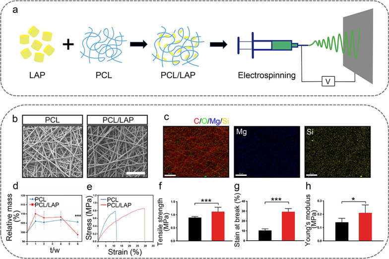
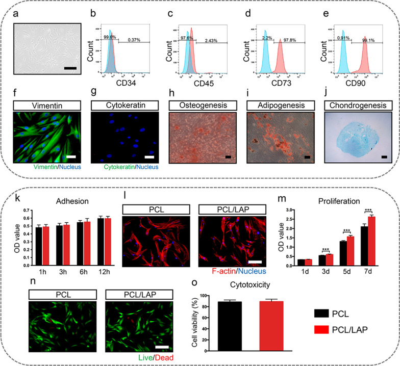
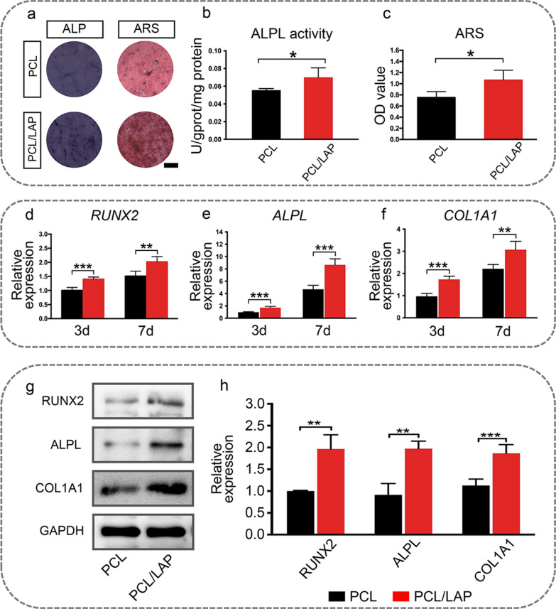
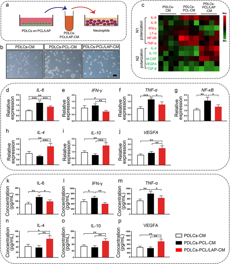
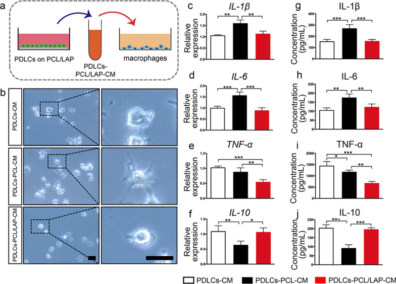
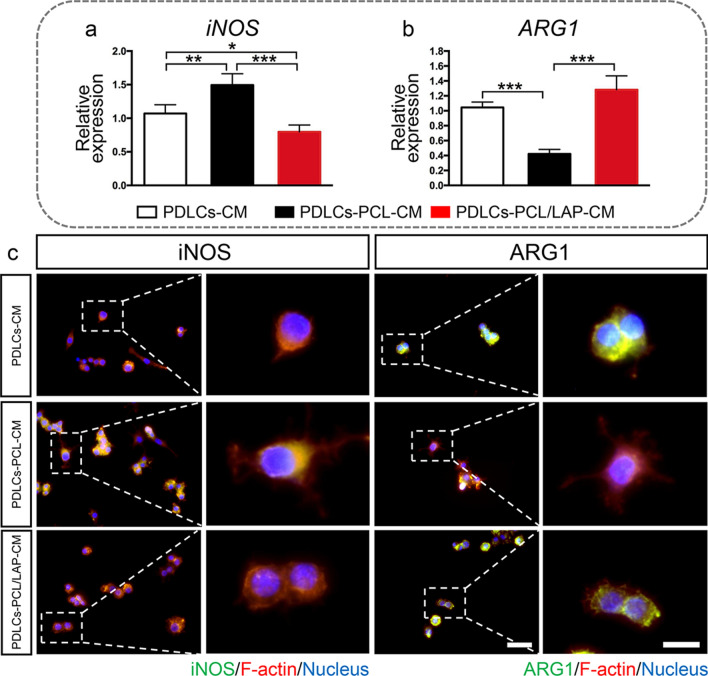
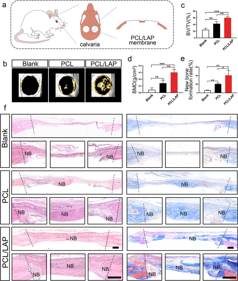
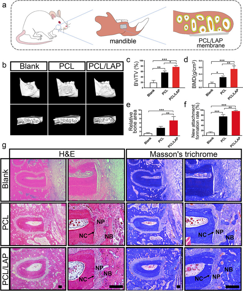
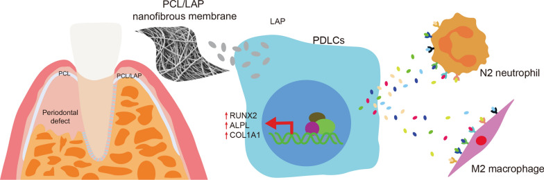
Although various new biomaterials have enriched the methods for periodontal regeneration, their efficacy is still controversial, and the regeneration of damaged support tissue in the periodontium remains challenging. Laponite (LAP) nanosilicate is a layered two-dimensional nanoscale, ultrathin nanomaterial with a unique structure and brilliant biocompatibility and bioactivity. This study aimed to investigate the effects of nanosilicate-incorporated PCL (PCL/LAP) nanofibrous membranes on periodontal ligament cells (PDLCs) in vitro and periodontal regeneration in vivo. A PCL/LAP nanofibrous membrane was fabricated by an electrospinning method. The characterization of PCL/LAP nanofibrous membrane were determined by scanning electron microscopy (SEM), energy dispersive spectrum of X-ray (EDS), inductively coupled plasma mass spectrometry (ICP-MS) and tensile test. The proliferation and osteogenic differentiation of PDLCs on the PCL/LAP nanofibrous membrane were evaluated. A PDLCs and macrophage coculture system was used to explore the immunomodulatory effects of the PCL/LAP nanofibrous membrane. PCL/LAP nanofibrous membrane was implanted into rat calvarial and periodontal defects, and the regenerative potential was evaluated by microcomputed topography (micro-CT) and histological analysis. The PCL/LAP nanofibrous membrane showed good biocompatibility and bioactivity. It enhanced the proliferation and osteogenic differentiation of PDLCs. The PCL/LAP nanofibrous membrane also stimulated anti-inflammatory and pro-remodeling N2 neutrophil formation, regulated inflammatory responses and induced M2 macrophage polarization by orchestrating the immunomodulatory effects of PDLCs. The PCL/LAP nanofibrous membrane promoted rat calvarial defect repair and periodontal regeneration in vivo. LAP nanosilicate-incorporated PCL membrane is capable of mediating osteogenesis and immunomodulation of PDLCs in vitro and accelerating periodontal regeneration in vivo. It could be a promising biomaterial for periodontal regeneration therapy.

Keywords: Immunomodulation; Nanosilicate; Osteogenesis; Periodontal ligament; Periodontal regeneration.


Conflict of interest statement

The authors declare that they have no known competing financial interests or personal relationships that could have appeared to influence the work reported in this paper

Figures

Fig. 1
Fig. 1
Characterization of PCL/LAP nanofibrous membranes. a Schematic diagram of the fabrication of PCL/LAP nanofibrous membrane. b SEM of PCL and PCL/LAP nanofibrous membranes. Scale bar = 20 μm. c EDS mapping analysis of PCL/LAP. Scale bar = 20 μm. d Degradation characteristics of PCL and PCL/LAP nanofibrous membranes. e The representative stress-strain curves of PCL and PCL/LAP nanofibrous membranes. f Tensile strength. g Strain at break. h Young’s modulus. Scale bar = 100 μm. *P ≤ 0.05, **P ≤ 0.01, ***P ≤ 0.001
Fig. 2
Fig. 2
Biocompatibility of PCL/LAP nanofibrous membranes. a PDLCs cultured from human PDL tissue. b-e Flow cytometry analysis of CD34, CD45, CD73 and CD90 in PDLCs. f-g Immunofluorecent staining of vimentin and cytokeratin in PDLCs. h ARS staining. i Oil red O staining. j Alcian blue staining. k Adhesion of PDLCs on PCL and PCL/LAP nanofibrous membranes. l Cytoskeleton staining of PDLCs on PCL and PCL/LAP nanofibrous membrane. m Proliferation of PDLCs on PCL and PCL/LAP nanofibrous membranes. n Live/dead staining and analysis of PDLCs on PCL and PCL/LAP nanofibrous membranes. Scale bar = 100 μm. *P ≤ 0.05, **P ≤ 0.01, ***P ≤ 0.001
Fig. 3
Fig. 3
PCL/LAP nanofibrous membrane enhances osteogenic differentiation of PDLCs. The PCL/LAP nanofibrous membrane enhances the osteogenic differentiation of PDLCs. a ALPL and ARS staining of PDLCs on PCL and PCL/LAP nanofibrous membranes. b ALPL activity of PDLCs on PCL and PCL/LAP nanofibrous membranes. c Quantification of ARS staining of PDLCs on PCL and PCL/LAP nanofibrous membranes. d-f Expression levels of osteogenic differentiation genes (RUNX2, ALPL and COL1A1) of PDLCs on PCL and PCL/LAP nanofibrous membranes. g-h Western blot analysis of osteogenic differentiation proteins (RUNX2, ALPL and COL1A1) of PDLCs on PCL and PCL/LAP nanofibrous membranes. Scale bar = 1 mm. *P ≤ 0.05; **P ≤ 0.01; ***P ≤ 0.001
Fig. 4
Fig. 4
Conditioned medium from PDLCs cultured on PCL/LAP nanofibrous membranes potential induces anti-inflammatory and pro-remodeling N2 neutrophils polarization. a Schematic illustration of neutrophils treated by conditioned medium from PDLCs. b Representative appearances of neutrophils treated with different condition medium. c Heat maps of gene expression levels in neutrophils treated with condition medium from PDLCs cultured on PCL/LAP nanofibrous membranes. d-g Pro-inflammatory gene (IL-6, IFN-γ, TNF-α and NF-κB) levels of neutrophils. h-i Anti-inflammatory gene (IL-4 and IL-10) expression of neutrophils. j Pro-remodeling gene (VEGFA) expression of neutrophils. k-m Pro-inflammatory cytokines (IL-6, IFN-γ and TNF-α) levels of neutrophils. n-o Anti-inflammatory cytokines (IL-4 and IL-10) levels of neutrophils. j Pro-remodeling cytokine (VEGFA) level of neutrophils. Scale bar = 30 μm. *P ≤ 0.05; **P ≤ 0.01; ***P ≤ 0.001
Fig. 5
Fig. 5
Conditioned medium from PDLCs cultured on PCL/LAP nanofibrous membranes inhibits macrophage inflammatory responses. a Schematic illustration of macrophages treated by conditioned medium from PDLCs. b Representative images of macrophages treated with conditioned medium from PDLCs cultured on PCL/LAP nanofibrous membranes. c-f Inflammatory gene (IL-1β, IL-6, TNF-α and IL-10) expression of macrophages treated with conditioned medium from PDLCs cultured on PCL/LAP membranes. gj Inflammatory cytokines (IL-1β, IL-6, TNF-α and IL-10) of macrophages treated with conditioned medium from PDLCs cultured on PCL/LAP membranes. Scale bar = 30 μm. *P ≤ 0.05; **P ≤ 0.01; ***P ≤ 0.001
Fig. 6
Fig. 6
Conditioned medium from PDLCs cultured on PCL/LAP nanofibrous membranes regulates macrophage polarization. a-b Decreased M1 polarization marker (iNOS) gene and increased M2 polarization marker (ARG1) gene levels of macrophages treated with condition medium from PDLCs cultured on PCL/LAP nanofibrous membrane. c Representative images of immunofluorescent staining iNOS levels and ARG1 levels in macrophages treated with conditioned medium from PDLCs cultured on PCL/LAP nanofibrous membranes. Scale bar = 50 μm. *P ≤ 0.05; **P ≤ 0.01; ***P ≤ 0.001
Fig. 7
Fig. 7
PCL/LAP nanofibrous membrane accelerates rat calvarial bone formation. a Schematic illustration of rat calvarial bone defect treated with PCL/LAP nanofibrous membrane. b Representative micro-CT reconstructed images of rat calvarial bone defects treated with PCL/LAP nanofibrous membranes. c-d BMD and BV/TV ratio of micro-CT data analysis. e Histological quantitative analysis of new bone formation rate. f Histological H&E- and Masson’s trichome-stained sections rat calvarial defects. NB, new bone. Scale bar = 200 μm. *P ≤ 0.05; **P ≤ 0.01; ***P ≤ 0.001
Fig. 8
Fig. 8
PCL/LAP nanofibrous membrane promotes rat mandibular periodontal defect regeneration. a Schematic illustration of rat periodontal defect treated with PCL/LAP nanofibrous membrane. b Representative micro-CT reconstructed images of periodontal defects treated with PCL/LAP nanofibrous membranes. c-d BV/TV ratio and BMD of Micro-CT data analysis. e-f Histological quantitative analysis of new bone area and new attachment formation rate. g Histological H&E- and Masson’s trichome-stained sections of rat periodontal defects. NC, new cementum; NP, new periodontal ligaments; NB, new bone. Scale bar = 200 μm. *P ≤ 0.05; **P ≤ 0.01; ***P ≤ 0.001
Fig. 9
Fig. 9
Graphical model of nanosilicate-functionalized PCL membrane improved periodontal regeneration by regulating PDLCs-mediated osteogenesis and immunomodulation in microenvironments after periodontal regeneration therapy

Similar articles

Cited by

References

    1. Slots J. Periodontitis: facts, fallacies and the future. Periodontol 2000. 2017;75:7–23. doi: 10.1111/prd.12221. - DOI - PubMed
    1. Florjanski W, Orzeszek S, Olchowy A, Grychowska N, Wieckiewicz W, Malysa A, Smardz J, Wieckiewicz M. Modifications of polymeric membranes used in guided tissue and bone regeneration. Polymers. 2019;11:782. doi: 10.3390/polym11050782. - DOI - PMC - PubMed
    1. Omar O, Elgali I, Dahlin C, Thomsen P. Barrier membranes: more than the barrier effect? J Clin Periodontol. 2019;46:103–23. doi: 10.1111/jcpe.13068. - DOI - PMC - PubMed
    1. Li J, Chen M, Wei X, Hao Y, Wang J. Evaluation of 3D-Printed polycaprolactone scaffolds coated with freeze-dried platelet-rich plasma for bone regeneration. Mater . 2017;10:831. doi: 10.3390/ma10070831. - DOI - PMC - PubMed
    1. Zhang L, Dong Y, Zhang N, Shi J, Zhang X, Qi C, Midgley A, Wang S. Potentials of sandwich-like chitosan/polycaprolactone/gelatin scaffolds for guided tissue regeneration membrane. Mater Sci Eng C Mater Biol Appl. 2020;109:110618. doi: 10.1016/j.msec.2019.110618. - DOI - PubMed