Review
. 2023 Jul 13;14(7):426.
doi: 10.1038/s41419-023-05969-9.

The Road from AKI to CKD: Molecular Mechanisms and Therapeutic Targets of Ferroptosis

Affiliations
Review

The Road from AKI to CKD: Molecular Mechanisms and Therapeutic Targets of Ferroptosis

Runzhi Guo et al. Cell Death Dis. .

Abstract

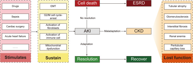
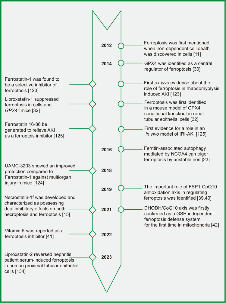
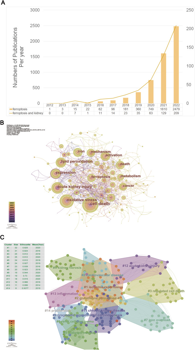
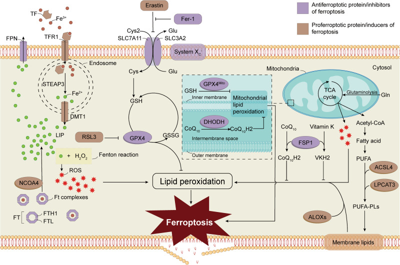
Acute kidney injury (AKI) is a prevalent pathological condition that is characterized by a precipitous decline in renal function. In recent years, a growing body of studies have demonstrated that renal maladaptation following AKI results in chronic kidney disease (CKD). Therefore, targeting the transition of AKI to CKD displays excellent therapeutic potential. However, the mechanism of AKI to CKD is mediated by multifactor, and there is still a lack of effective treatments. Ferroptosis, a novel nonapoptotic form of cell death, is believed to have a role in the AKI to CKD progression. In this study, we retrospectively examined the history and characteristics of ferroptosis, summarized ferroptosis's research progress in AKI and CKD, and discussed how ferroptosis participates in regulating the pathological mechanism in the progression of AKI to CKD. Furthermore, we highlighted the limitations of present research and projected the future evolution of ferroptosis. We hope this work will provide clues for further studies of ferroptosis in AKI to CKD and contribute to the study of effective therapeutic targets to prevent the progression of kidney diseases.


Conflict of interest statement

The authors declare no competing interests.

Figures

Fig. 1
Fig. 1. Evolution after kidney damage.
Adverse stimuli such as infection, nephrotoxic drugs, and hypoperfusion induce a series of reactions, including the immune system response and cell metabolic reprogramming in the kidney. Continuous and severe stimulation can lead to cell death, organ failure, and end-stage renal disease (ESRD). Moreover, different responses to cell repair determine different prognoses. Some cells are repaired, regenerated, and cured, while maladaptive cells undergo renal tubular atrophy, renal interstitial fibrosis, and glomerulosclerosis through AKI to CKD and gradually enter the ESRD stage. EMT epithelial-mesenchymal transition, AKI acute kidney injury, CKD chronic kidney disease, ESRD end-stage renal disease.
Fig. 2
Fig. 2. Timeline of the development of key discoveries in ferroptosis regulatory mechanisms and inhibitors.
This figure depicts a timeline of significant discoveries in the regulation of ferroptosis and the creation of inhibitors for it. The timeline emphasizes crucial events, such as the initial identification of ferroptosis in cells, in animal models, and in kidney disease. The purpose of this figure is to provide a comprehensive summary of the advancements made in comprehending and treating ferroptosis from different perspectives.
Fig. 3
Fig. 3. Bibliometric analyses of ferroptosis in the kidney.
Data were extracted from the Web of Science database, and bibliometric analysis was performed using CiteSpace, a web-based Java application for data analysis and visualization. The keywords finally identified as follows: 1st: [TS = ferroptosis or TS = ferroptotic) and ((PY = (2012–2022)) AND DT = (Article OR Review)) AND LA = (English). 2nd: [TS = ferroptosis or TS = ferroptotic) and (((((TS = (kidney)) OR TS = (renal)) OR TS = (nephr*)) OR TS = (Glomer*)) OR TS = (podocyte)) OR TS = (“Proximal tubular”) and ((PY= (2012–2022)) AND DT = (Article OR Review)) AND LA = (English) (A) Number of publications per year; (B) Keywords co-occurrence analysis; (C) Keywords cluster view.
Fig. 4
Fig. 4. Schematic diagram of the primary regulatory mechanisms associated with ferroptosis.
Lipid peroxidation is necessary for ferroptosis in individual cells, in which iron-induced ROS burst and the decrease of antioxidation are required. Several molecular mechanisms were reported to regulate the occurrence of ferroptosis, such as the system Xc-/GPX4 antioxidant axis, iron regulons NCOA4 and DMT1. Furthermore, ACSL4, LPCAT3, and ALOXs induced ferroptosis by influencing the levels of cellular lipid peroxides. ROS reactive oxygen species, Cys2 cystine, Cys cysteine, Glu glutamate, GSH glutathione, Gln glutamine, TCA cycle tricarboxylic acid cycle, DHODH dihydroorotate dehydrogenase, CoQ coenzyme Q, GPX4 glutathione peroxidase 4, PUFAs polyunsaturated fatty acids, ACSL4 acyl-CoA synthetase long-chain family member 4, LPCAT3 lysophosphatidylcholine acyltransferase 3, FSP1, ferroptosis suppressor protein 1, ALOX arachidonate 5-lipoxygenase, TF transferrin, TFR1 transferrin receptor 1, DMT1 divalent metal transporter 1, Ft ferritin, FTH1 ferritin heavy chain, FTL ferritin light chain, NCOA4 nuclear receptor coactivator 4, Fer-1 ferrostatin-1, FPN ferroportin, LIP labile iron pool, SLC7A11 solute carrier family 7 member 11, SLC3A2 solute carrier family 3 member 2, STEAP3 six-transmembrane epithelial antigen of the prostate 3.
Fig. 5
Fig. 5. The crosstalk between inflammation and ferroptosis.
As a major representative cell of the inflammatory response, macrophages are activated in response to PAMPs and DAMPs after intense noxious stimulation. The inflammatory state may have led to an energy disorder, increased the level of LIP, and attenuated the beneficial effect of antioxidants, which further triggered ferroptosis. Cell death leads to the release of DAMPs which stimulate inflammation. Thus a positive feedback loop forms and ultimately leads to organ injury.

References

    1. Al-Jaghbeer M, Dealmeida D, Bilderback A, Ambrosino R, Kellum JA. Clinical decision support for in-hospital AKI. J Am Soc Nephrol. 2018;29:654–60. doi: 10.1681/ASN.2017070765. - DOI - PMC - PubMed
    1. Susantitaphong P, Cruz DN, Cerda J, Abulfaraj M, Alqahtani F, Koulouridis I, et al. World incidence of AKI: a meta-analysis. Clin J Am Soc Nephrol. 2013;8:1482–93. doi: 10.2215/CJN.00710113. - DOI - PMC - PubMed
    1. Ronco C, Bellomo R, Kellum JA. Acute kidney injury. Lancet (Lond, Engl) 2019;394:1949–64. doi: 10.1016/S0140-6736(19)32563-2. - DOI - PubMed
    1. Sato Y, Takahashi M, Yanagita M. Pathophysiology of AKI to CKD progression. Semin Nephrol. 2020;40:206–15. doi: 10.1016/j.semnephrol.2020.01.011. - DOI - PubMed
    1. Global, regional, and national burden of chronic kidney disease, 1990-2017: a systematic analysis for the Global Burden of Disease Study 2017. Lancet (London, England). 2020;395:709–33. - PMC - PubMed

Publication types