. 2023 Jul 13;14(1):4160.
doi: 10.1038/s41467-023-39819-1.

Atomic structure of a nudivirus occlusion body protein determined from a 70-year-old crystal sample

Affiliations

Atomic structure of a nudivirus occlusion body protein determined from a 70-year-old crystal sample

Jeremy R Keown et al. Nat Commun. .

Abstract

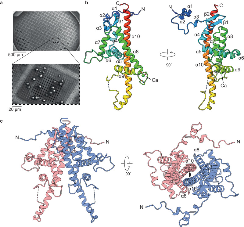
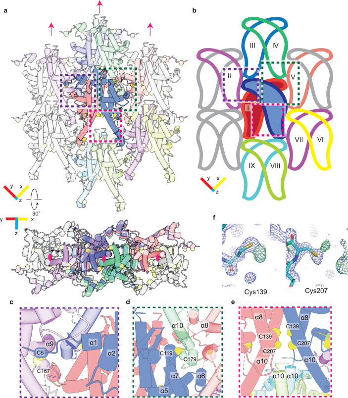
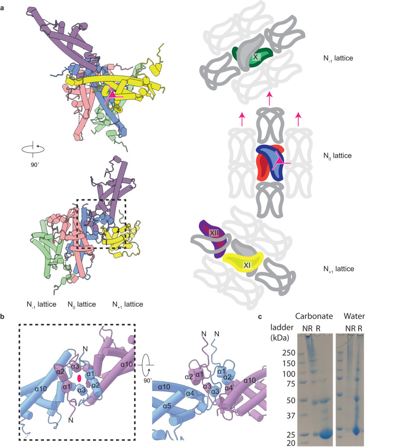
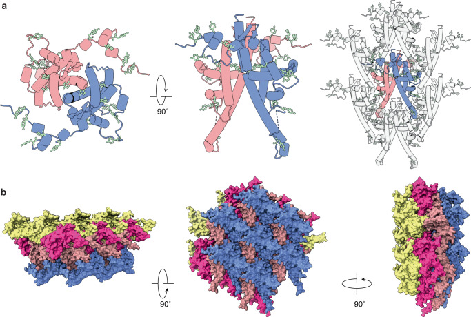
Infectious protein crystals are an essential part of the viral lifecycle for double-stranded DNA Baculoviridae and double-stranded RNA cypoviruses. These viral protein crystals, termed occlusion bodies or polyhedra, are dense protein assemblies that form a crystalline array, encasing newly formed virions. Here, using X-ray crystallography we determine the structure of a polyhedrin from Nudiviridae. This double-stranded DNA virus family is a sister-group to the baculoviruses, whose members were thought to lack occlusion bodies. The 70-year-old sample contains a well-ordered lattice formed by a predominantly α-helical building block that assembles into a dense, highly interconnected protein crystal. The lattice is maintained by extensive hydrophobic and electrostatic interactions, disulfide bonds, and domain switching. The resulting lattice is resistant to most environmental stresses. Comparison of this structure to baculovirus or cypovirus polyhedra shows a distinct protein structure, crystal space group, and unit cell dimensions, however, all polyhedra utilise common principles of occlusion body assembly.


Conflict of interest statement

The authors declare no competing interests.

Figures

Fig. 1
Fig. 1. Structure of the ToNV polyhedrin.
a Scanning electron micrograph of native ToNV occlusion bodies prepared for diffraction experiments. Images have been cropped but are otherwise unedited. b A single polyhedrin molecule coloured from the N-terminus (blue) to the C-terminus (red) and annotated with secondary structure features. A calcium ion is shown (green). The dashed line shows the missing loop 171–174. c The dimeric unit (red and blue chains) of the OB lattice are shown in two orientations. The two-fold symmetric axis is indicated by the black ellipse.
Fig. 2
Fig. 2. The 2D lattice packs tightly and is maintained through disulfide bond formation.
a, b Display of the 2D lattice, the reference polyhedrin (blue) and the polyhedrin with which it makes contact are shown in various colours. The two polyhedrin molecules which form the repeating unit are shown without transparency (a) and with fill colour (b). Molecules which do not contact the reference molecule are in grey. Pink arrows and ellipses indicate the 2-fold crystallographic axis. c, d Detailed insets showing the residues which form the asymmetric disulfide bonds. e C139/C207 are partially oxidised cysteine residues positioned near to the unordered C-terminus of neighbouring molecules. f Additional electron density observed at C139/C207. The 2FOFc map at 1σ (blue) and FO – Fc at 3σ are shown.
Fig. 3
Fig. 3. Electrostatic, hydrophobic, and domain swapping maintains interactions between lattice sheets.
a Polyhedrin molecules which form major interfaces between the sheets of the polyhedra. The reference polyhedrin dimer (blue/red), with polyhedrin from the sheets in front (yellow/purple) and behind (green) are shown. A simplified scheme (right side) showing the relative arrangement of polyhedrin around the three-fold crystallographic axis (pink triangle), two-fold axis (pink arrow), and the change in direction of each sequential sheet in the lattice. b Detailed view of the domain swapping interactions annotated with secondary structure names. The crystallographic 2-fold axis is shown (pink ellipse). c SDS-PAGE of recombinant crystals treated with NaH2CO3 or water under reducing (R) or non-reducing (NR) conditions. Samples were prepared twice and a representative image shown.
Fig. 4
Fig. 4. The occlusion body is impermeable and poised for dissolution.
a Two polyhedrin molecules (red/blue) which form the dimeric repeating building block and position of tyrosine residues (pale green). Arrangement of the residues on the dimer and their resulting position in the lattice are shown. b Two sheets of the lattice (red/blue and pink/yellow) are shown from three orientations demonstrating that the assembled crystal does not contain any open channels.

References

    1. Chiu E, Coulibaly F, Metcalf P. Insect virus polyhedra, infectious protein crystals that contain virus particles. Curr. Opin. Struct. Biol. 2012;22:234–240. doi: 10.1016/j.sbi.2012.02.003. - DOI - PubMed
    1. Glaser RW, Chapman JW. The nature of the polyhedral bodies found in insects. Biol. Bull. 1916;30:367–390. doi: 10.2307/1536363. - DOI
    1. Rohrmann GF, Pearson MN, Bailey TJ, Becker RR, Beaudreau GS. N-terminal polyhedrin sequences and occludedBaculovirus evolution. J. Mol. Evol. 1981;17:329–333. doi: 10.1007/BF01734354. - DOI - PubMed
    1. Rohrmann GF. Polyhedrin structure. J. Gen. Virol. 1986;67:1499–1513. doi: 10.1099/0022-1317-67-8-1499. - DOI - PubMed
    1. Arella M, Lavallée C, Belloncik S, Furuichi Y. Molecular cloning and characterization of cytoplasmic polyhedrosis virus polyhedrin and a viable deletion mutant gene. J. Virol. 1988;62:211–217. doi: 10.1128/jvi.62.1.211-217.1988. - DOI - PMC - PubMed

Publication types