. 2023 Jul 13;14(1):4187.
doi: 10.1038/s41467-023-39908-1.

Biophysical ordering transitions underlie genome 3D re-organization during cricket spermiogenesis

Affiliations

Biophysical ordering transitions underlie genome 3D re-organization during cricket spermiogenesis

Guillermo A Orsi et al. Nat Commun. .

Abstract

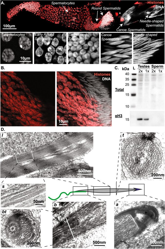
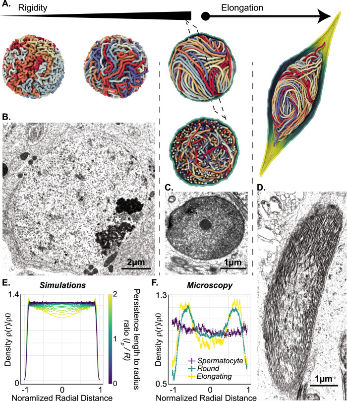
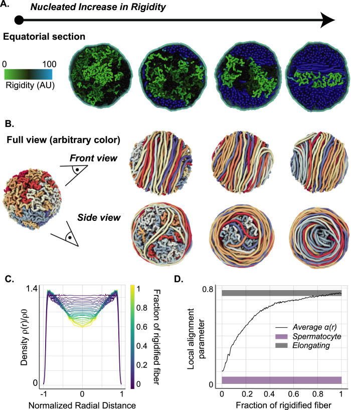
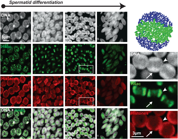
Spermiogenesis is a radical process of differentiation whereby sperm cells acquire a compact and specialized morphology to cope with the constraints of sexual reproduction while preserving their main cargo, an intact copy of the paternal genome. In animals, this often involves the replacement of most histones by sperm-specific nuclear basic proteins (SNBPs). Yet, how the SNBP-structured genome achieves compaction and accommodates shaping remain largely unknown. Here, we exploit confocal, electron and super-resolution microscopy, coupled with polymer modeling to identify the higher-order architecture of sperm chromatin in the needle-shaped nucleus of the emerging model cricket Gryllus bimaculatus. Accompanying spermatid differentiation, the SNBP-based genome is strikingly reorganized as ~25nm-thick fibers orderly coiled along the elongated nucleus axis. This chromatin spool is further found to achieve large-scale helical twisting in the final stages of spermiogenesis, favoring its ultracompaction. We reveal that these dramatic transitions may be recapitulated by a surprisingly simple biophysical principle based on a nucleated rigidification of chromatin linked to the histone-to-SNBP transition within a confined nuclear space. Our work highlights a unique, liquid crystal-like mode of higher-order genome organization in ultracompact cricket sperm, and establishes a multidisciplinary methodological framework to explore the diversity of non-canonical modes of DNA organization.


Conflict of interest statement

The authors declare no competing interests.

Figures

Fig. 1
Fig. 1. Coiled organization of chromatin fibers in Gryllus bimaculatus sperm.
A Overview of spermiogenesis in Gryllus bimaculatus. Confocal microscopy image of a single follicle from an adult testis stained for DNA (white) and histones (red). Multiple cysts are visible in which male germ cells differentiate synchronously. From our estimations, the premeiotic cysts contain 512 primary spermatocytes and post-meiotic cysts must then contain about 2048 spermatids. By analogy to Drosophila, we distinguish successive stages in spermiogenesis including spermatocytes, round spermatids, canoe (elongating) spermatids, and needle-shaped spermatids. *: a polyploid somatic nucleus. B, C Sperm chromatin undergoes a Histone-SNBP transition. Confocal image showing early elongating (left side) and late elongating (right side) spermatids stained for DNA (white) and histones (red) confirms massive histone eviction during spermatid elongation (B). Western blot of protein extracts from testes and sperm revealing whole proteins and histone H3 confirms undetectable histone levels in mature gametes (C). L: ladder, 2x 1x: two-fold dilution of protein extract. D Chromatin organizes as coiled fibers in spermatids. Electron microscopy images show different sections of spermatids around the canoe stage. A longitudinal section (l) of the spermatid nucleus shows chromatin fibers in alignment with the antero-posterior axis. A sagittal section (s) of individual chromatin fibers reveals a tubular internal structure. At the basal end (b, bt), an overlap between the nucleus and flagellum is observed. Note that images in (b) and (bt) represent longitudinal and transversal sections of this structure. Bending of chromatin fibers is observed at the apical end (a). Approximately 200 individualized fibers are distinguished in a transversal section (t).
Fig. 2
Fig. 2. Polymer modeling of rigid fiber dynamics recapitulates features of chromatin localization in sperm.
A Chromatin re-organization via a uniform, homogeneous increase in local fiber rigidity in simulated models. Polymer simulations of the stiffening process resulting in a yarn-ball-like state, which is relatively disordered and prevents the establishment of a coherent, homogeneous direction of fiber alignment upon the onset of nuclear elongation. BD Chromatin re-organization during spermiogenesis in vivo. Electron microscopy images of spermatocytes (B), round (C), and elongating (D) spermatids show progressive reorganization of chromatin by fiber individualization and alignment. E Simulated radial monomer density profiles, normalized by the mean nuclear fiber concentration ρ0, for different persistence-length-to-radius ratios lp/R. Distance is normalized to nucleus radius. Note the growing fiber accumulation at the nuclear periphery as rigidification progresses and lp/R increases. F Experimental density profiles, as computed from TEM images of spermatocytes (purple), round spermatids (green), and elongating spermatids (yellow)(see Methods). Distance is normalized to nucleus radius. Error bars denote the standard error of the mean, and were evaluated using O(10) independent micrographs taken at the spermatocyte (B), round (C), and elongating (D) developmental stages. In B, ‘ * ‘ indicates a heterochromatin mass.
Fig. 3
Fig. 3. Nucleated chromatin rigidification results in a spool-like organization.
A Chromatin re-organization via a nucleated, in cis propagation of fiber rigidification. Fiber segments undergoing gradual stiffening are marked in green, denoting a putative peak in acetylation activity. Fully-rigidified segments are depicted in blue, representing SNBP-based chromatin. Unmodified chromatin regions are rendered in black. B Same as Fig. 3a in full-fiber view. The segregation of rigidified chromatin segments into a toroidal structure at the nuclear periphery is clearly visible, and leads to the formation of a spool-like ordered structure towards the late round spermatid stage. C Simulated radial monomer density profiles at different rigidified fiber fractions. Distance is normalized to nucleus radius. Nucleated rigidification results in a significantly more pronounced peripheral accumulation of chromatin than the simple uniform stiffening process (Fig. 2E), although the mechanical properties of the fully-rigidified fiber are identical in both cases. D Radially-averaged local alignment parameter α(r), as computed from the simulations (black line) and evaluated from TEM images at the spermatocyte (purple box) and elongating stage (gray box). The width of the experimental bars represents the standard error of the mean (see “Methods” section).
Fig. 4
Fig. 4. Histone acetylation patterns in vivo are consistent with the rigidification dynamics predicted by our model.
Confocal images of spermatids at four early, consecutive stages in spermiogenesis stained for DNA (white), pan-acetylated histone H4 (H4ac, green), and total histones (red). H4ac appears as punctate nuclear foci that cluster at the nucleus center, subsequently spreading as two waves directed towards opposite nuclear poles (see arrowheads in magnified panels on the right). At this stage, DNA is enriched in the complementary nuclear space surrounding the central region (arrows). This organization is consistent with our simulations. Early elongating spermatids (right panels) show dispersed and weaker H4ac and histone signals.
Fig. 5
Fig. 5. Cellular polarization precedes chromatin orientation.
A, B Round spermatids feature nascent flagella and acrosomes. A: Confocal microscopy images of testes stained for DNA (white) and αTubulin (green) show nascent flagellar microtubules in early spermatids. B Electron microscopy image of a round spermatid cyst showing multiple nuclei (left). The boxed region is zoomed in the right panel. Chromatin is homogeneously enriched at the nuclear periphery, flanked by nascent flagellar (arrow) and acrosomal (arrowhead) structures, indicating that cellular antero-posterior poles were defined before chromatin became oriented. C In polymer simulations, nuclear elongation drives the spontaneous reorientation of the chromatin spool axis perpendicular to the antero-posterior line, and leads to the distinct unidirectional alignment of the chromatin fiber coils. D Elongating spool organization observed in simulations and in vivo. Transversal sections of nuclei during elongation in polymer simulations (left) and in electron microscopy images of elongating spermatids (right) show a similar configuration whereby the vestigial spool axis perpendicular to the elongation axis is still apparent. E Alignment parameter α computed through the bulk of the nuclear interior (see Methods), as a function of the applied stretching force F. As in Fig. 3D, bars correspond to the standard error of the mean from experimental measurements from EM images at the elongating stage (gray box).
Fig. 6
Fig. 6. Chromatin twists around the nucleus axis in late spermiogenesis.
A, B DNA twisting in late spermatids. Single Molecule Localization Microscopy (SMLM) images of late canoe (A) and fully mature (B) spermatids labeled with an HMSiR-Hoechst probe to reveal DNA with nanoscopic resolution. Top panels show the epifluorescence image; middle and bottom panels show SMLM images and close in. DNA twists around the nucleus axis over several turns. Boxed regions are zoomed in bottom panels. C In simulated data, a geometric twist imposes an additional extensional strain on the peripheral chromatin fibers, which leads to an increased degree of lateral compaction upon mechanical relaxation. Chromatin configuration and density in simulated polymers before and after relaxation recapitulate that observed in vivo by electron microscopy in late canoe (bottom left) and mature (bottom right) spermatids respectively. D Evidence for contacts between the flagellum and chromatin. Electron microscopy transversal sections of the basal end of canoe spermatids reveal a proteinaceous structure connecting the flagellar axoneme and chromatin fibers (full arrow). Close to its organizing center, the flagellum fully penetrates the nucleus and is fully embedded by chromatin (dashed arrow). E Whole-nucleus looping in late spermatids. Confocal microscopy images of late needle-shaped spermatid cells in testes labeled for DNA with DAPI, showing occasional curling. The boxed region is zoomed in the right panel.
Fig. 7
Fig. 7. Disulfide bridging maintains nuclear compaction and shaping.
A Nuclear decondensation is modeled by the release of applied extensional forces, and leads to a rapid relaxation towards a spherical envelope shape via a transient, corkscrew-like intermediary morphology. B Chromatin is readily decondensed upon treatment with reducing agent DTT. Epifluorescence images of mature spermatids isolated from female spermatheca, labeled for DNA and exposed to 25 mM DTT. Chromatin decondensation pattern is consistent with simulated data and suggests that protamine disulfide bridges maintain the needle-like nuclear morphology.

References

    1. O’Donnell L. Mechanisms of spermiogenesis and spermiation and how they are disturbed. Spermatogenesis. 2014;4:e979623. doi: 10.4161/21565562.2014.979623. - DOI - PMC - PubMed
    1. Lüpold S, Pitnick S. Sperm form and function: what do we know about the role of sexual selection? Reproduction. 2018;155:R229–R243. doi: 10.1530/REP-17-0536. - DOI - PubMed
    1. Lee JD, 4th, Allen MJ, Balhorn R. Atomic force microscope analysis of chromatin volumes in human sperm with head-shape abnormalities. Biol. Reprod. 1997;56:42–49. doi: 10.1095/biolreprod56.1.42. - DOI - PubMed
    1. Wyrobek AJ, Meistrich ML, Furrer R, Bruce WR. Physical characteristics of mouse sperm nuclei. Biophys. J. 1976;16:811–825. doi: 10.1016/S0006-3495(76)85730-X. - DOI - PMC - PubMed
    1. Ward WS, Coffey DS. DNA packaging and organization in mammalian spermatozoa: comparison with somatic cells. Biol. Reprod. 1991;44:569–574. doi: 10.1095/biolreprod44.4.569. - DOI - PubMed

Publication types