

Caspase activation in tumour-infiltrating lymphocytes is associated with lymph node metastasis in oral squamous cell carcinoma
- PMID: 37443405
- PMCID: PMC10772935
- DOI: 10.1002/path.6145
Caspase activation in tumour-infiltrating lymphocytes is associated with lymph node metastasis in oral squamous cell carcinoma
Abstract
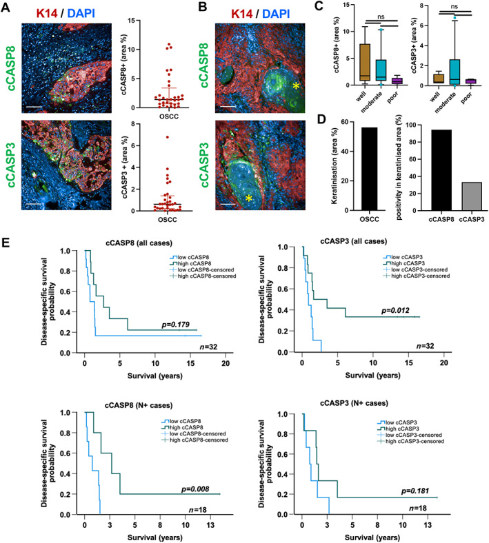
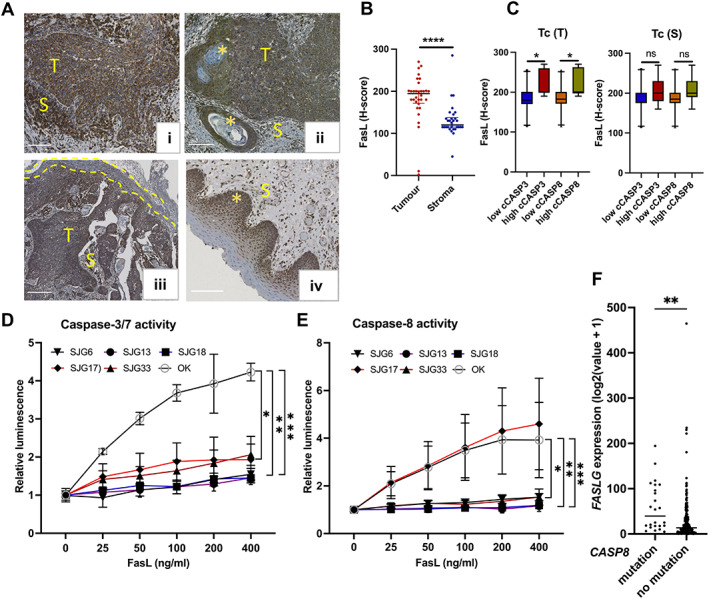
Oral squamous cell carcinomas (OSCCs) are genetically heterogeneous and exhibit diverse stromal and immune microenvironments. Acquired resistance to standard chemo-, radio-, and targeted therapies remains a major hurdle in planning effective treatment modalities for OSCC patients. Since Caspase 8 (CASP8) is frequently mutated in OSCCs, we were interested to explore a potential interaction between tumour-infiltrating lymphocytes (TILs) and CASP8 activation using high-content image analysis of human tumour (n = 32) sections. Despite the lymphocyte-rich tumour microenvironment, we observed lower activation of CASP8 (0-10% of tumour area) and its downstream effector CASP3 (0-6%) in tumours than in normal oral epithelium. Conversely, we found apoptosis was high for all the lymphocyte subtypes examined (38-52% of lymphocytes within tumour islands). Tumours with higher Fas ligand (FasL) expression had a significantly higher proportion of cleaved CASP3/8 positive cytotoxic T cells within the tumour islands (p = 0.05), and this was associated with the presence of lymph node metastatic disease [odds ratio: 1.046, 95% confidence interval (1.002-1.091), p = 0.039]. Our finding of extensive activation of the extrinsic pathway of apoptosis in TILs, together with evidence of higher FasL in CASP8 mutated tumours, may be useful in predicting the course of disease in individual patients. © 2023 The Authors. The Journal of Pathology published by John Wiley & Sons Ltd on behalf of The Pathological Society of Great Britain and Ireland.
Keywords: CASPASE8; OSCC; T cell exhaustion; apoptosis; apoptotic T lymphocytes; cytotoxic T lymphocytes; immune evasion; lymph node metastasis; tumour-infiltrating lymphocytes (TILs).
© 2023 The Authors. The Journal of Pathology published by John Wiley & Sons Ltd on behalf of The Pathological Society of Great Britain and Ireland.
Figures


Similar articles
-
Role Of Cd8+ Tumour-Infiltrating Lymphocytes In Predicting Regional Lymph Node Metastasis In Lip And Oral Cavity Squamous Cell Carcinoma.J Ayub Med Coll Abbottabad. 2023 Apr-Jun;35(2):288-293. doi: 10.55519/JAMC-02-11654. J Ayub Med Coll Abbottabad. 2023. PMID: 37422823
-
A digital score of tumour-associated stroma infiltrating lymphocytes predicts survival in head and neck squamous cell carcinoma.J Pathol. 2022 Feb;256(2):174-185. doi: 10.1002/path.5819. Epub 2021 Nov 24. J Pathol. 2022. PMID: 34698394
-
Effects of FasL expression in oral squamous cell cancer.Asian Pac J Cancer Prev. 2013;14(1):281-5. doi: 10.7314/apjcp.2013.14.1.281. Asian Pac J Cancer Prev. 2013. PMID: 23534738
-
The prognostic role of tumour-infiltrating lymphocytes in oral squamous cell carcinoma: A meta-analysis.J Oral Pathol Med. 2019 Oct;48(9):788-798. doi: 10.1111/jop.12927. Epub 2019 Aug 7. J Oral Pathol Med. 2019. PMID: 31323145 Review.
-
Tumor-infiltrating ICOS+ Effector Regulatory T-Cells in Oral Squamous Cell Carcinoma as a Promising Biomarker for Prognosis and 'Hot' Tumor.Anticancer Res. 2022 May;42(5):2383-2393. doi: 10.21873/anticanres.15717. Anticancer Res. 2022. PMID: 35489733 Review.
Cited by
-
Tumor microenvironment in oral squamous cell carcinoma.Front Immunol. 2024 Dec 18;15:1485174. doi: 10.3389/fimmu.2024.1485174. eCollection 2024. Front Immunol. 2024. PMID: 39744628 Free PMC article. Review.
-
Exploring beyond Common Cell Death Pathways in Oral Cancer: A Systematic Review.Biology (Basel). 2024 Feb 6;13(2):103. doi: 10.3390/biology13020103. Biology (Basel). 2024. PMID: 38392321 Free PMC article. Review.
-
Caspase-8-dependent autophagy regulates neutrophil infiltration in oral squamous cell carcinoma.Proc Natl Acad Sci U S A. 2024 Dec 10;121(50):e2406944121. doi: 10.1073/pnas.2406944121. Epub 2024 Dec 3. Proc Natl Acad Sci U S A. 2024. PMID: 39625985 Free PMC article.
References
-
- Sung H, Ferlay J, Siegel RL, et al. Global cancer statistics 2020: GLOBOCAN estimates of incidence and mortality worldwide for 36 cancers in 185 countries. CA Cancer J Clin 2021; 71: 209–249. - PubMed
-
- Cancer Research UK . Head and Neck Cancers Survival Statistics. [Accessed 19 May 2022]. Available from: https://www.cancerresearchuk.org/health‐professional/cancer‐statistics/s....
-
- Brandwein‐Gensler M, Teixeira MS, Lewis CM, et al. Oral squamous cell carcinoma: histologic risk assessment, but not margin status, is strongly predictive of local disease‐free and overall survival. Am J Surg Pathol 2005; 29: 167–178. - PubMed
Publication types
MeSH terms
Substances
Grants and funding
LinkOut - more resources
Full Text Sources
Medical
Research Materials
Miscellaneous
