

Inter-Rater Agreement for Diagnosing Adenomyosis Using Magnetic Resonance Imaging and Transvaginal Ultrasonography
- PMID: 37443587
- PMCID: PMC10341351
- DOI: 10.3390/diagnostics13132193
Inter-Rater Agreement for Diagnosing Adenomyosis Using Magnetic Resonance Imaging and Transvaginal Ultrasonography
Abstract
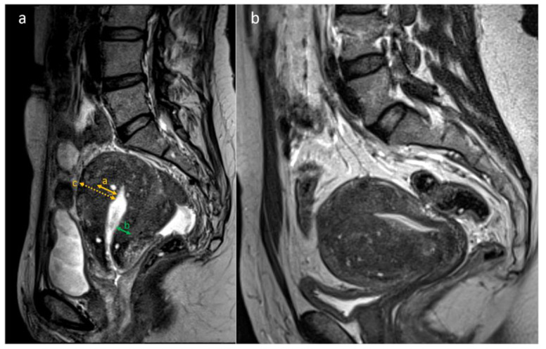
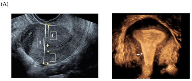
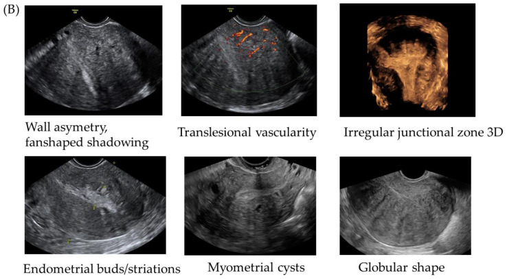
Our aim was to compare the inter-rater agreement about transvaginal ultrasonography (TVS) with magnetic resonance imaging (MRI) with regard to diagnosing adenomyosis and for assessing various predefined imaging features of adenomyosis, in the same set of women. The study cohort included 51 women, prospectively, consecutively recruited based on a clinical suspicion of adenomyosis. MRIs and TVS videoclips and 3D volumes were retrospectively assessed by four experienced radiologists and five experienced sonographers, respectively. Each rater subjectively evaluated the presence or absence of adenomyosis, as well as imaging features suggestive of adenomyosis. Fleiss kappa (κ) was used to reflect inter-rater agreement for categorical data, and the intraclass correlation coefficient (ICC) was used to reflect the reliability of quantitative data. Agreement between raters for diagnosing adenomyosis was higher for TVS than for MRI (κ = 0.42 vs. 0.28). MRI had a higher inter-rater agreement in assessing wall asymmetry, irregular junctional zone (JZ), and the presence of myometrial cysts, while TVU had a better agreement for assessing globular shape. MRI showed a moderate to good reliability for measuring the JZ (ICC = 0.57-0.82). For TVS, the JZ was unmeasurable in >50% of cases, and the remaining cases had low reliability (ICC = -0.31-0.08). We found that inter-rater agreement for diagnosing adenomyosis was higher for TVS than for MRI, despite the fact that MRI showed a higher inter-rater agreement in most specific features. Measurements of JZ in the coronal plane with 3D TVS were unreliable and thus unlikely to be useful for diagnosing adenomyosis.
Keywords: adenomyosis; inter-rater agreement; magnetic resonance imaging; ultrasonography.
Conflict of interest statement
The authors declare no conflict of interest. The funders had no role in the design of the study; in the collection, analyses, or interpretation of data; in the writing of the manuscript; or in the decision to publish the results.
Figures
Similar articles
-
Inter-rater agreement in the diagnosis of adenomyosis by 2- and 3-dimensional transvaginal ultrasonography.J Ultrasound Med. 2019 Mar;38(3):657-666. doi: 10.1002/jum.14735. Epub 2018 Sep 4. J Ultrasound Med. 2019. PMID: 30182497
-
Transvaginal sonographic features of diffuse adenomyosis in 18-30-year-old nulligravid women without endometriosis: association with symptoms.Ultrasound Obstet Gynecol. 2015 Dec;46(6):730-6. doi: 10.1002/uog.14834. Ultrasound Obstet Gynecol. 2015. PMID: 25728241
-
Adenomyosis: three-dimensional sonographic findings of the junctional zone and correlation with histology.Ultrasound Obstet Gynecol. 2011 Apr;37(4):471-9. doi: 10.1002/uog.8900. Ultrasound Obstet Gynecol. 2011. PMID: 21433167
-
Uterine junctional zone and adenomyosis: comparison of MRI, transvaginal ultrasound and histology.Ultrasound Obstet Gynecol. 2023 Jul;62(1):42-60. doi: 10.1002/uog.26117. Epub 2023 Jun 1. Ultrasound Obstet Gynecol. 2023. PMID: 36370446 Review.
-
Role of transvaginal sonography and magnetic resonance imaging in the diagnosis of uterine adenomyosis.Fertil Steril. 2018 Mar;109(3):389-397. doi: 10.1016/j.fertnstert.2018.01.024. Fertil Steril. 2018. PMID: 29566851 Review.
Cited by
-
How Reproducible Are the Ultrasound Features of Adenomyosis Defined by the Revised MUSA Consensus?J Clin Med. 2025 Jan 13;14(2):456. doi: 10.3390/jcm14020456. J Clin Med. 2025. PMID: 39860462 Free PMC article.
References
-
- Bird C.C., McElin T.W., Manalo-Estrella P. The elusive adenomyosis of the uterus—Revisited. Am. J. Obstet. Gynecol. 1972;112:583–593. - PubMed
-
- Cockerham A.Z. Adenomyosis: A Challenge in Clinical Gynecology. J. Midwifery Womens Health. 2012;57:212–220. - PubMed
-
- Pinzauti S., Lazzeri L., Tosti C., Centini G., Orlandini C., Luisi S., Zupi E., Exacoustos C., Petraglia F. Transvaginal sonographic features of diffuse adenomyosis in 18–30-year-old nulligravid women without endometriosis: Association with symptoms. Ultrasound Obstet. Gynecol. 2015;46:730–736. - PubMed
-
- Bazot M., Darai E. Role of transvaginal sonography and magnetic resonance imaging in the diagnosis of uterine adenomyosis. Fertil. Steril. 2018;109:389–397. - PubMed
Grants and funding
LinkOut - more resources
Full Text Sources
