. 2023 Jul 5;13(13):2282.
doi: 10.3390/diagnostics13132282.

Performance Analysis of Segmentation and Classification of CT-Scanned Ovarian Tumours Using U-Net and Deep Convolutional Neural Networks

Affiliations

Performance Analysis of Segmentation and Classification of CT-Scanned Ovarian Tumours Using U-Net and Deep Convolutional Neural Networks

Ashwini Kodipalli et al. Diagnostics (Basel). .

Abstract

Difficulty in detecting tumours in early stages is the major cause of mortalities in patients, despite the advancements in treatment and research regarding ovarian cancer. Deep learning algorithms were applied to serve the purpose as a diagnostic tool and applied to CT scan images of the ovarian region. The images went through a series of pre-processing techniques and, further, the tumour was segmented using the UNet model. The instances were then classified into two categories-benign and malignant tumours. Classification was performed using deep learning models like CNN, ResNet, DenseNet, Inception-ResNet, VGG16 and Xception, along with machine learning models such as Random Forest, Gradient Boosting, AdaBoosting and XGBoosting. DenseNet 121 emerges as the best model on this dataset after applying optimization on the machine learning models by obtaining an accuracy of 95.7%. The current work demonstrates the comparison of multiple CNN architectures with common machine learning algorithms, with and without optimization techniques applied.

Keywords: DenseNet; Dice score; Jaccard score; ResNet; UNet; VGG 16; convolutional neural networks; ovarian tumours.


Conflict of interest statement

The authors declare no conflict of interest.

Figures

Figure 1
Figure 1
U-Net model architecture for segmentation of benign and malignant tumours.
Figure 2
Figure 2
Flow of the implementation.
Figure 3
Figure 3
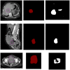
Benign dataset. Towards the (left) is the input image, the (middle) is the label and the (right) is the segmented image.
Figure 3
Figure 3
Benign dataset. Towards the (left) is the input image, the (middle) is the label and the (right) is the segmented image.
Figure 4
Figure 4
Malignant dataset. Towards the (left) is the input image, the (middle) is the label and the (right) is the segmented image.
Figure 5
Figure 5
Mean plot of benign and malignant performance.
Figure 6
Figure 6
Dice score comparison between benign and malignant tumours.
Figure 7
Figure 7
Jaccard score comparison between benign and malignant tumours.
Figure 8
Figure 8
Diagrammatic representation of the performance of the CNN models.
Figure 9
Figure 9
Diagrammatic representation of the comparison of the results with tuning techniques.

Similar articles

Cited by

References

    1. Labidi-Galy S.I., Treilleux I., Goddard-Leon S., Combes J.-D., Blay J.-Y., Ray-Coquard I., Caux C., Bendriss-Vermare N. Plasmacytoid dendritic cells infiltrating ovarian cancer are associated with poor prognosis. Oncoimmunology. 2012;1:380–382. doi: 10.4161/onci.18801. - DOI - PMC - PubMed
    1. Siegel R.L., Miller K.D., Wagle N.S., Jemal A. Cancer statistics. CA Cancer J. Clin. 2023;73:17–48. doi: 10.3322/caac.21763. - DOI - PubMed
    1. Jung Y., Kim T., Han M.R., Kim S., Kim G., Lee S., Choi Y.J. Ovarian tumour diagnosis using deep convolutional neural networks and a denoising convolutional autoencoder. Sci. Rep. 2022;12:17024. doi: 10.1038/s41598-022-20653-2. - DOI - PMC - PubMed
    1. Wang X., Li H., Zheng P. Automatic Detection and Segmentation of Ovarian Cancer Using a Multitask Model in Pelvic CT Images. Oxidative Med. Cell. Longev. 2022;2022:6009107. doi: 10.1155/2022/6009107. - DOI - PMC - PubMed
    1. Mahmood F., Borders D., Chen R.J., Mckay G.N., Salimian K.J., Baras A., Durr N.J. Deep Adversarial Training for Multi-Organ Nuclei Segmentation in Histopathology Images. IEEE Trans. Med. Imaging. 2019;39:3257–3267. doi: 10.1109/TMI.2019.2927182. - DOI - PMC - PubMed

LinkOut - more resources