Review
. 2023 Jun 23;12(13):1699.
doi: 10.3390/cells12131699.

Could a Non-Cellular Molecular Interactome in the Blood Circulation Influence Pathogens' Infectivity?

Affiliations
Review

Could a Non-Cellular Molecular Interactome in the Blood Circulation Influence Pathogens' Infectivity?

Eugenio Hardy et al. Cells. .

Abstract

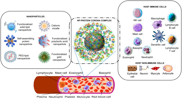
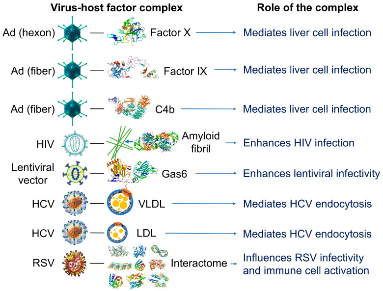
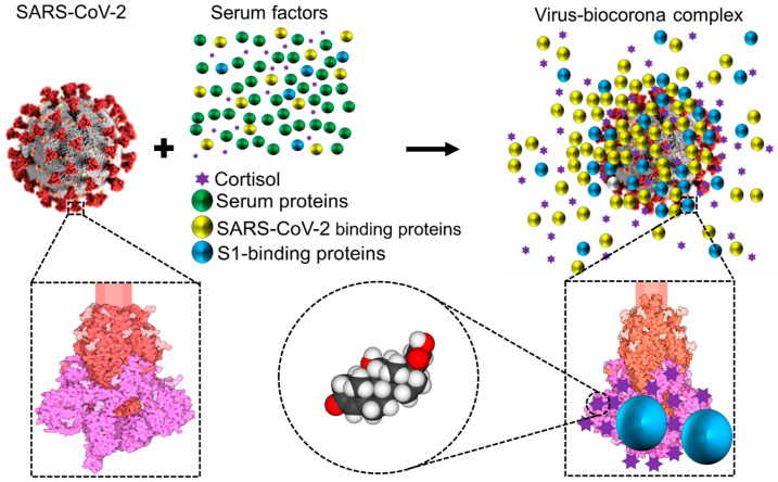
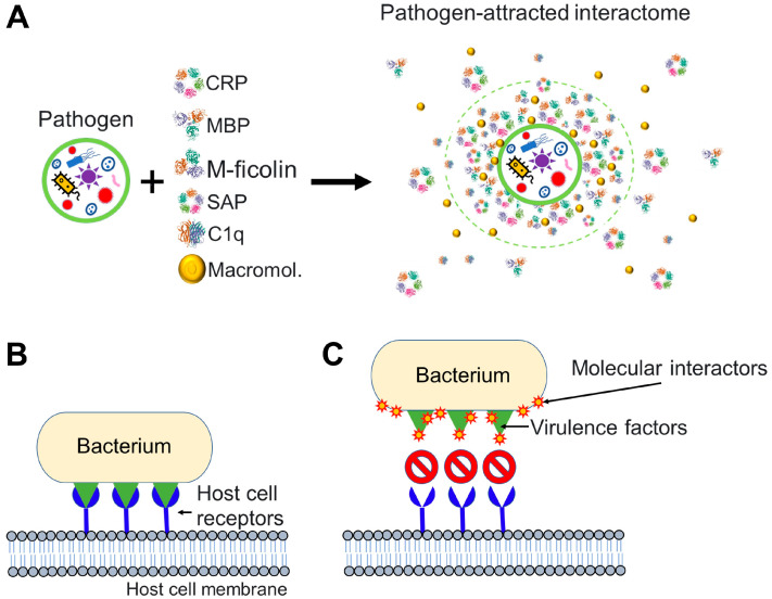
We advance the notion that much like artificial nanoparticles, relatively more complex biological entities with nanometric dimensions such as pathogens (viruses, bacteria, and other microorganisms) may also acquire a biomolecular corona upon entering the blood circulation of an organism. We view this biomolecular corona as a component of a much broader non-cellular blood interactome that can be highly specific to the organism, akin to components of the innate immune response to an invading pathogen. We review published supporting data and generalize these notions from artificial nanoparticles to viruses and bacteria. Characterization of the non-cellular blood interactome of an organism may help explain apparent differences in the susceptibility to pathogens among individuals. The non-cellular blood interactome is a candidate therapeutic target to treat infectious and non-infectious conditions.

Keywords: bacteria; glucocorticoid; innate immunity; nanoparticles; protein corona; virus.


Conflict of interest statement

The authors declare no conflict of interest.

Figures

Figure 1
Figure 1
The nanoparticle protein corona interacts with host cells. Synthetic nanoparticles (on the left) enter the bloodstream, where they interact with proteins (a proteomic non-cellular blood interactome) to form a nanoparticle (NP)–protein corona complex (middle panel). The protein corona gives the synthetic nanoparticle a biological identity that mediates interactions with innate immune cells and non-immune cells (right panel). The protein corona can also effectively hide surface ligands, reducing the interaction with intended target cell receptors [30]. The protein corona–host cell interaction activates biological responses, including innate immune recognition and clearance by phagocytic cells, adaptive immune response, therapeutic activity, and toxic reactions such as thrombocyte activation, hemolysis, and excessive pattern-recognition receptor-mediated inflammation [17,30,31,32,33,34]. Immune cells may recognize exposed corona proteins as antigens and trigger undesirable immune reactions [30,35]. Image files used to partly generate the figure are licensed under https://creativecommons.org/licenses/by/4.0 (I53-50 nanoparticle vaccine schematic.png by J. Marcandalli et al. (accessed on 1 April 2023), Vaccines-09-00065-g001.webp by M.D. Buschmann et al. (2021), Delivery methods for HIV mRNA vaccine.png by Z. Mu et al. (accessed on 5 April 2023), and Mechanisms_by_which_bacteria_target_tumors.svg by M. T-Q. Duong et al. (accessed on 7 April 2023)), https://creativecommons.org/licenses/by-sa/4.0 (DNA-AuNP0022.jpg by Shpetrosko (accessed on 4 April 2023), Giemsa_Stain_Macrophage_Illustration.png by Noah Smith (accessed on 18 April 2023), Final_stem_cell_differentiation_(1).svg by Haileyfournier (accessed on 14 April 2023), 3FFN_background_removed.png by Pronchik (accessed on 6 April 2023), and Antibody_IgG1_structure.png by Tokenzero (accessed on 8 April 2023)), https://creativecommons.org/licenses/by/3.0 (Blausen 0909 WhiteBloodCells.png by Blausen.com staff (accessed on 14 April 2023)), https://creativecommons.org/licenses/by-sa/3.0 (SolidLipidNanoparticle.jpg by Andrea Trementozzi (accessed on 8 April 2023), Cardiovascular system-Lymphopoiesis-NK cell—Smart-Servier.png by Laboratoires Servier (accessed on 13 April 2023), and Protein_CFB_PDB_1dle.png, Protein_TF_PDB_1a8e.png, Protein_C3_PDB_1c3d.pngtructure of the C3 protein and Protein_CP_PDB_1kcw.png, all by Emw (accessed on 11 April 2023)), https://creativecommons.org/licenses/by/2.0 (Mast Cell (30107399584).jpg by NIAID (accessed on 18 April 2023)), and https://creativecommons.org/publicdomain/zero/1.0/ (PDB_1ao6_EBI.jpg by Jawahar Swaminathan, C5.png by Pietro Roversi, Antithrombin_monomer.jpeg by K. Murphy, and Lymphocyte, Mast cell, Eosinophil, Basophil, Neutrophil, Platelet, Monocyte and Red blood cell, all by Sarbasst Braian), via Wikimedia Commons. Abbreviations: PEG, polyethyleneglycol; NK cell, natural killer cell.
Figure 2
Figure 2
Examples of reported interactions involving viruses and components of the blood interactome (left panel). Impact of the resulting complexes on viral infectivity and host immune response (right panel). Adenovirus (Ad), a 90–100 https://en.wikipedia.org/wiki/Nanometernm virus composed of double-stranded DNA, an icosahedral nucleocapsid, and three major capsid proteins (hexon, penton, and fiber), attracts and interacts with blood factors including coagulation factors (factor IX and factor X) and complements C4B [56,57,58,59]. Human immunodeficiency virus (HIV), a single-stranded, positive-sense ribonucleic acid-containing, spherical (100–120 nm), enveloped virus that also contains a capsid core and proteins (e.g., gp120 and gp41) embedded in the lipid bilayer, attracts and interacts with fibril-forming prostatic acid phosphatase fragments [60]. Lentiviral vectors, a type of retroviruses originated from human immunodeficiency viruses, attract and interact with a gamma-carboxyglutamic acid domain-containing protein called Gas6 (growth arrest-specific 6). Lentiviral vectors pseudotyped with envelope proteins from various sources, including the Ross River virus, Sindbis virus, and baculovirus, induce this interaction [61]. Hepatitis C virus (HCV), a small, enveloped virus of 55–65 nm in diameter, consisting of positive-sense single-stranded RNA and glycoproteins (e.g., E1 and E2) embedded in the lipid bilayer, attracts and interacts with lipoproteins such as low-density lipoproteins (LDL) and very low-density lipoproteins (VLDL) [62]. Respiratory syncytial virus (RSV), an enveloped, spherical, and sometimes filamentous (150 nm) virus with a single-stranded negative-sense RNA, a capsid core, and glycoproteins (e.g., such as F, G, and SH proteins) embedded in the lipid bilayer, attracts and interacts with hundreds of host proteins depending on the extracellular biologically fluid (e.g., human plasma and bronchoalveolar lavage fluid) [63]. In all cases, the resulting acquired corona serves as the surface for actual contact with host cells and may influence the ability of viruses to activate and enter the host cells. Instead of just one protein component, the combination of corona components that are enriched on the viral surface affects viral infectivity [63]. The image files that were used to generate part of the illustration are licensed under https://creativecommons.org/licenses/by-sa/4.0 (Adenovirus_3D_schematic.png (accessed on 5 March 2023) and HI-virion-structure en.svg (accessed on 1 March 2023) by T. Splettstoesser and HCV.png by P. Znamenskiy (accessed on 4 March 2023)), https://creativecommons.org/licenses/by/3.0 (595768.fig.001.jpg by S.S. Bawage et al. (accessed on 11 March 2023)), https://creativecommons.org/licenses/by-sa/3.0 (GAPDH_with_labels.png by Vossman (accessed on 5 March 2023), Actin_with_ADP_highlighted.png by T. Splettstoesser (accessed on 3 March 2023), and Protein_F10_PDB_1c5m.png (accessed on 10 March 2023), Protein ACPP PDB 1cvi.png (accessed on 8 March 2023), Protein_GAS6_PDB_1h30.png (accessed on 12 March 2023), Protein_TUBA1A_PDB_1ffx.png (accessed on 17 March 2023), Protein C3 PDB 1c3d.png (accessed on 15 March 2023), Protein_C4A_PDB_1hzf.png (accessed on 11 March 2023), Protein_HSPA1A_PDB_1hjo.png (accessed on 16 March 2023), and Protein_FGB_PDB_1fza.png (accessed on 14 March 2023) all by Emw), and https://creativecommons.org/publicdomain/zero/1.0/ (Lentiviral_vector.png by P. Znamenskiy, PDB_1pfx_EBI.jpg by J. Swaminathan and MSD staff, PBB_Protein_APOA1_image.jpg by ProteinBoxBot at English Wikipedia, and Lactoferrin.png by Lijealso), via Wikimedia Commons. PDB entry 5JTW was used for C4b [64].
Figure 3
Figure 3
Schematic representation showing that while circulating in the extracellular environment viral particles may undergo biophysical transformations, such as becoming decorated by blood components leading to a biocorona—analogous to what has been described for nanoparticles. This notion is illustrated and has been documented for the case of SARS-CoV-2 [6,73]. Notice that the ‘yellow and blue’ (virus-binding) proteins are enriched on the virus surface, whereas the ‘green’ (non-binding) proteins are not. S1 is colored in magenta, S2 in red, and glycosylation is shown in lighter shades. Representation of the SARS-CoV Spike protein (https://cdn.rcsb.org/pdb101/motm/246/246-SARSCoV2_Spike-6crz_6vxx3.tif), which is shown magnified in this illustration, was obtained from David Goodsell (https://doi.org/10.2210/rcsb_pdb/mom_2020_6), licensed under CC BY 4.0 (https://creativecommons.org/licenses/by/4.0/). The original illustration was created using PDB entries 6crz [74] and 6vxx [75]. Abbreviations: SARS-CoV-2, severe acute respiratory syndrome-coronavirus-2; S, spike.
Figure 4
Figure 4
Pathogen interactions with molecules of the host can affect pathogen infectivity. (A) Pathogen interacts with molecular factors from the bloodstream and attracts an interactome to its surface. Components of the pathogen-attracted interactome may include pentraxins such as C-reactive protein (CRP) and serum amyloid P, collectins such as mannose-binding lectin (MBL), ficolins such as M-ficolin, complement molecules such as the complement component 1q (C1q), and many other macromolecules (Macromol.) including endogenous cortisol [6,73] and proteins and peptides. (B,C) Particular case where the invading pathogen is a bacterium. (B) An invading bacterium interacts with host cell receptors (in blue) through virulence factors (in green). Virulence factors are molecules that facilitate host invasion by bacterial cells. (C) Molecular interactome components are drawn to pathogenic bacteria and successfully prevent the interaction between the virulence factors of the bacterium and host cell receptors. To generate the figure, we used the PDB entry 1GNH for CRP [81], PDB entry 1RTM for MBP [82], PDB entry 2D39 for M-ficolin [83], PDB entry 1SAC for serum amyloid P [84], and PDB entry 1PK6 for C1q [85].

Similar articles

References

    1. Kumar H., Kawai T., Akira S. Pathogen Recognition by the Innate Immune System. Int. Rev. Immunol. 2011;30:16–34. doi: 10.3109/08830185.2010.529976. - DOI - PubMed
    1. Janeway C.A., Medzhitov R. Innate Immune Recognition. Annu. Rev. Immunol. 2002;20:197–216. doi: 10.1146/annurev.immunol.20.083001.084359. - DOI - PubMed
    1. Medzhitov R., Janeway C. Innate Immune Recognition: Mechanisms and Pathways. Immunol. Rev. 2000;173:89–97. doi: 10.1034/j.1600-065X.2000.917309.x. - DOI - PubMed
    1. Medzhitov R., Janeway C. Innate Immunity. N. Engl. J. Med. 2000;343:338–344. doi: 10.1056/NEJM200008033430506. - DOI - PubMed
    1. Paludan S.R., Pradeu T., Masters S.L., Mogensen T.H. Constitutive Immune Mechanisms: Mediators of Host Defence and Immune Regulation. Nat. Rev. Immunol. 2020;21:137–150. doi: 10.1038/s41577-020-0391-5. - DOI - PMC - PubMed

Publication types

LinkOut - more resources