. 2023 Jun 27;12(13):1733.
doi: 10.3390/cells12131733.

Pleiotrophin and the Expression of Its Receptors during Development of the Human Cerebellar Cortex

Affiliations

Pleiotrophin and the Expression of Its Receptors during Development of the Human Cerebellar Cortex

Margarita Belem Santana-Bejarano et al. Cells. .

Abstract

During embryonic and fetal development, the cerebellum undergoes several histological changes that require a specific microenvironment. Pleiotrophin (PTN) has been related to cerebral and cerebellar cortex ontogenesis in different species. PTN signaling includes PTPRZ1, ALK, and NRP-1 receptors, which are implicated in cell differentiation, migration, and proliferation. However, its involvement in human cerebellar development has not been described so far. Therefore, we investigated whether PTN and its receptors were expressed in the human cerebellar cortex during fetal and early neonatal development. The expression profile of PTN and its receptors was analyzed using an immunohistochemical method. PTN, PTPRZ1, and NRP-1 were expressed from week 17 to the postnatal stage, with variable expression among granule cell precursors, glial cells, and Purkinje cells. ALK was only expressed during week 31. These results suggest that, in the fetal and neonatal human cerebellum, PTN is involved in cell communication through granule cell precursors, Bergmann glia, and Purkinje cells via PTPRZ1, NRP-1, and ALK signaling. This communication could be involved in cell proliferation and cellular migration. Overall, the present study represents the first characterization of PTN, PTPRZ1, ALK, and NRP-1 expression in human tissues, suggesting their involvement in cerebellar cortex development.

Keywords: ALK; NRP-1; RPTPZ1; cerebellum; development; human; pleiotrophin.


Conflict of interest statement

The authors declare no conflict of interest.

Figures

Figure 1
Figure 1
Histological changes in cerebellar cortex in human development. (AF) Hematoxylin and eosin staining of fetal cerebellar and neonatal samples; (G) timeline of morphological changes in the cerebellar cortex. Dark blue—fetal samples. Light blue—neonatal sample. EGL—external granular layer; IGL—internal granular layer; IL—internal layer; ML—molecular layer; PCL—Purkinje cell layer. The scale bar in image A is 20 µm and applies to all microphotographs. The red arrows show PCs.
Figure 2
Figure 2
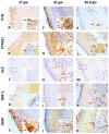
Representative image of PTN and the expression of its receptors in the human cerebellar cortex, from 17 gw to 36.6 gw. (AD) The expression of PTN, PTPRZ1, and NRP-1 are low; ALK is negative at 17 gw. (E) GFAP expression is similar to PTN staining; (FI) the expression of PTN, PTPRZ1, ALK, and NRP-1 is high at 31 gw; (J,O) GFAP pattern expression is similar to PTPRZ1 expression; (KN) the expression of PTN and NRP-1 is limited to PCs, PTPRZ1 has diffuse distribution, and ALK is negative. The scale bar in image (A) is 50 µm and applies to all microphotographs.
Figure 3
Figure 3
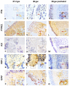
Representative image of PTN and the expression of its receptors in human cerebellar cortex from 37.4 gw to neonatal stage. (AD) The expression of PTN and NRP-1 is limited to PCs, PTPRZ1 has a high intensity, ALK is negative at 37.4 gw; (E,J,O) GFAP pattern expression is similar to PTPRZ1 distribution; (FI) PTN has a low intensity in PCs, NRP-1 has a moderate intensity in PCs, PTPRZ1 shows diffuse distribution, ALK is negative at 38 gw; (KN) PTN expression is located in EGL, NRP-1 has a low intensity in PCs, PTPRZ1 has a high intensity in all cerebellar cortex layers; ALK is negative at the neonatal stage. The scale bar in image (A) is 50 µm and applies to all microphotographs.
Figure 4
Figure 4
Schematic illustration of the spatiotemporal expression of PTN and its receptors from 17 gw to the neonatal sample of the human cerebellar cortex. EGL—external granular layer; IGL—internal granular layer; IL—internal layer; ML—molecular layer; PCL—Purkinje cell layer. PTN (yellow). PTPRZ1 (green). ALK (purple). NRP-1 (pink). Negative (black). The triangle size is related to protein level expression. Created on BioRender.com.

References

    1. Haldipur P., Bharti U., Alberti C., Sarkar C., Gulati G., Iyengar S., Gressens P., Mani S. Preterm Delivery Disrupts the Developmental Program of the Cerebellum. PLoS ONE. 2011;6:e23449. doi: 10.1371/journal.pone.0023449. - DOI - PMC - PubMed
    1. Larsell O. The Development of the Cerebellum in Man in Relation to Its Comparative Anatomy. J. Comp. Neurol. 1947;87:85–129. doi: 10.1002/cne.900870203. - DOI - PubMed
    1. Rakic P., Sidman R.L. Histogenesis of Cortical Layers in Human Cerebellum, Particularly the Lamina Dissecans. J. Comp. Neurol. 1970;139:473–500. doi: 10.1002/cne.901390407. - DOI - PubMed
    1. Amore G., Spoto G., Ieni A., Vetri L., Quatrosi G., Di Rosa G., Nicotera A.G. A Focus on the Cerebellum: From Embryogenesis to an Age-Related Clinical Perspective. Front. Syst. Neurosci. 2021;15:646052. doi: 10.3389/fnsys.2021.646052. - DOI - PMC - PubMed
    1. Millen K.J., Gleeson J.G. Cerebellar Development and Disease. Curr. Opin. Neurobiol. 2008;18:12–19. doi: 10.1016/j.conb.2008.05.010. - DOI - PMC - PubMed

Publication types

MeSH terms

LinkOut - more resources