Review
. 2023 Jun 28;12(13):1740.
doi: 10.3390/cells12131740.

Cardiovascular Functions of Ena/VASP Proteins: Past, Present and Beyond

Affiliations
Review

Cardiovascular Functions of Ena/VASP Proteins: Past, Present and Beyond

Peter M Benz et al. Cells. .

Abstract

Actin binding proteins are of crucial importance for the spatiotemporal regulation of actin cytoskeletal dynamics, thereby mediating a tremendous range of cellular processes. Since their initial discovery more than 30 years ago, the enabled/vasodilator-stimulated phosphoprotein (Ena/VASP) family has evolved as one of the most fascinating and versatile family of actin regulating proteins. The proteins directly enhance actin filament assembly, but they also organize higher order actin networks and link kinase signaling pathways to actin filament assembly. Thereby, Ena/VASP proteins regulate dynamic cellular processes ranging from membrane protrusions and trafficking, and cell-cell and cell-matrix adhesions, to the generation of mechanical tension and contractile force. Important insights have been gained into the physiological functions of Ena/VASP proteins in platelets, leukocytes, endothelial cells, smooth muscle cells and cardiomyocytes. In this review, we summarize the unique and redundant functions of Ena/VASP proteins in cardiovascular cells and discuss the underlying molecular mechanisms.

Keywords: Ena/VASP proteins; actin dynamics; angiogenesis; cardiomyocyte contraction; conducted vasodilation; endothelial barrier function; gap junction assembly; leukocyte infiltration and polarization; receptor trafficking; smooth muscle cell relaxation.


Conflict of interest statement

The authors declare no conflict of interest.

Figures

Figure 1
Figure 1
Actin-polymerization-driven processes in eukaryotic cells. Directed actin polymerization is the driving force for many cellular processes that shape and move cellular membranes. These processes include cell migration and contraction, endocytosis, phagocytosis and intracellular vesicle trafficking (A), as well as the assembly and disassembly of cell-cell junctions and cell-matrix adhesions (BF). Regulated by a plethora of actin binding proteins, including the Ena/VASP proteins (green), dynamic actin polymerization generates higher order actin networks and membrane protrusions (magenta). These include the rod-shaped actin stress fibers required for cell contraction; rod-shaped filopodia and microspikes, and branched lamellipodia crucial for cell migration; as well as the branched actin networks in endocytic/phagocytic membrane invaginations and actin comet tails regulating intracellular vesicle transport (A). In endothelial cells (BF), the formation of the cortical actin ring stabilizes tight and adherens junctions, thereby limiting paracellular permeability and vascular leakage (B,D). Inflammatory mediators and vasoactive substances induce the transition of the cortical acting ring into contractile stress fibers, which destabilize cell-cell junctions and pull opposing plasma membranes apart to increase paracellular permeability (C,E,F). In confluent ECV304 endothelial cells, actin dynamics form a cortical actin ring that lines the cytoplasmic face of cell-cell contacts (D, arrowheads). Following complexation of extracellular calcium by EGTA (E 15 min, F 30 min), perijunctional actin rings disassemble, focal adhesion associated stress fibers form (E,F white arrows), and interendothelial junctions open (E,F black arrows). Scale bars: 20 μm.
Figure 2
Figure 2
Subcellular localization of Ena/VASP proteins at sites of high actin turnover. (A,B) VASP in transfected Ena/VASP-deficient fibroblasts (MVD7) localized predominantly to the leading edge of lamellipodia and tips of microspikes within the lamellipodia (A, magnified views), and at the tips of filopodia (B). (C) In stably adherent ECV304 endothelial cells, VASP is clearly seen at integrin-based focal adhesions, which anchor actin stress fibers to the extracellular matrix. However, VASP also decorates stress fibers themselves in a punctate pattern (see magnified views). (D) Subcellular distribution of Mena in freshly isolated vascular smooth muscle cells (VSMCs) from mouse aorta. (E) In human umbilical vein endothelial cells (HUVECs), VASP colocalizes with actin and the gap junction protein connexin 40 (Cx40) at interendothelial junctions. Scale bars: 20 μm; magnified views: 10 μm.
Figure 3
Figure 3
Domain organization and phosphorylation sites of Ena/VASP proteins along with their binding partners and associated functions. EVH1: Ena/VASP homology 1, PRR: proline-rich region, LERER: low complexity region harbouring LERER repeats (unique to Mena), EVH2: Ena/VASP homology 2, GAB: G-actin binding site, FAB: F-actin binding site, CoCo: coiled coil motif required for tetramerization. Numbering according to the predominant human protein isoforms. Serine, threonine and tyrosine phosphorylation sites and the respective kinases are also indicated. Except for VASP S239 (which is present in Mena but not in EVL) and T278 (which is unique to VASP), all phosphorylation sites are structurally/functionally conserved in the Ena/VASP protein family (color-coded circles).
Figure 4
Figure 4
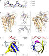
EVH1 and SH3 domain-mediated protein-protein interactions of Ena/VASP proteins. (A) Ball/stick model of a typical polyproline type II (PPII) helix as determined by X-ray diffraction (modified from pdb ID: 1FYN). PPII are left-handed helices that can be represented as triangular prism. Three consecutive prolines account for one turn of the PPII helix and occupy a different edge of the prism. (B) Ribbon diagram of the Mena EVH1 domain in complex with a FPPPP core peptide ligand. The overall fold of EVH1 domains is a compact, parallel β-sandwich capped along one side by a long α-helix. The highly conserved triad of surface-exposed aromatic sidechains, Y16, W23, and F77 (F79 in VASP), come together in the 3-dimensional structure of the domain to form an aromatic cluster, which provides a hydrophobic docking site for the proline-rich peptide ligands (modified from pdb ID: 1evh). (C) Ribbon model of chicken α-spectrin SH3 domain as determined by X-ray diffraction (pdb ID: 1SHG) in a hypothetical complex with a proline-rich ligand (PPPVPPRV, pdb ID: 1CKB). The SH3 domain is a compact β-barrel made of five antiparallel β-strands (βA-βE) that are connected (from N- to C-terminus) by the RT, n-Src, and distal loop, and by a 310 helix, respectively. β-Strands βA and βE, and β-strands βB-βD form two tightly packed anti-parallel β sheets that are shown in blue or yellow, respectively. The β-barrel is shown in two different orientations related by a 90° rotation.
Figure 5
Figure 5
Comparison of the proline-rich regions from human EVL, VASP, and Mena. Prolines are shown in red, glycines preceding a proline-stretch in blue. The preferred PKA phosphorylation site in VASP, S157 (green), is located in close proximity to the GP5 motifs. The PRR of Mena is the largest, spanning 64 amino acids, followed by VASP with 50, and EVL with only 25. EVL contains a single GP8 motif, VASP a triple GP5 motif, and Mena a GP6 and GP9 motif.
Figure 6
Figure 6
Role of Ena/VASP proteins in endothelial cell repulsion from ephrin ligands. HUVECs, treated with control (A,B) or Mena/VASP (C,D) siRNA, were seeded on alternating 50 μm stripes with clustered ephrin-B2 (labeled with Cy3 fluorescent dye) or control and imaged by time-lapse microcopy. Representative phase contrast images of cells approximately two hours (A,C, magnified view) or five hours (B,D) after seeding are shown; ephrin-B2 stripes are indicated by red overlays. Please note the cytoskeletal collapse in control siRNA-transfected HUVECs on ephrin-B2 stripes, resulting in dot-like structures and membrane ruffles (A, white and black arrows, respectively), whereas no cytoskeletal collapse was observed in Mena/VASP siRNA transfected cells on ephrin-B2 stripes (C, compare lamellipodia indicated by black and white arrowheads, respectively). Figure modified from [127].
Figure 7
Figure 7
Proposed model how VASP-deficiency increases monocyte recruitment, macrophage polarization and vascular repair after ischemia. (A) VASP-deficiency drives monocyte infiltration into the ischemic muscle through decreased CCR2 receptor internalization. The latter is likely caused by reduced β-arrestin-2 signaling and/or reduced actin dynamics that drive membrane trafficking. VASP-deficiency impairs AMPK phosphorylation (activation), which drives STAT1-dependent macrophage polarization and CCL2 release. Together, the two mechanisms synergistically increase leukocyte recruitment into the ischemic tissue, which in turn drives angiogenesis, arteriogenesis and vascular repair. (B) Sequence alignment of rat (Rn), mouse (Mm), human (Hs), and bull (Bt) PP1-R6 protein sequence. The putative VASP EVH1 motif is highlighted in red. The EVH1 core binding motif and the second, high-affinity EVH1 binding motif of listeria (Lm) ActA are shown for comparison (x, any amino acid; Φ, hydrophobic amino acid). (C,D) Proposed model how VASP deficiency impairs AMPK phosphorylation in macrophages. Phosphorylation of AMPK at Thr-172 is induced by AMP/ADP and the upstream protein kinases LKB1, CaMKKβ and TAK1. Dephosphorylation of AMPK at Thr172 is regulated by protein phosphatases PP1, PP2A and PP2C. (C) In wild-type cells, VASP-binding to PP1-R6/PP1 complex limits the PP1-dependent de-phosphorylation of AMPK. In the absence of VASP, AMPK dephosphorylation by the PP1-R6/PP1 complex is increased. (D) In macrophages, AMPK activation drives phagocytosis and inhibits JAK-STAT and NF-κB signaling, thereby limiting the expression of pro-inflammatory cytokines including TNFα and IL-1β. Conversely, impaired AMPK activity in VASP-deficient macrophages increases the STAT1-mediated pro-inflammatory phenotype and limits phagocytosis. Figure modified from [63].
Figure 8
Figure 8
Critical role of VASP in supporting the conducted vasodilation along the vessel wall. (A) Schematic diagram showing how conducted vasodilations can be studied in the microcirculation in vivo. A glass micropipette is positioned in close proximity to a second- or third-order arteriole (resting diameter ~10–15 μm) of the cremaster muscle. Locally confined application of acetylcholine is used to elicit endothelial hyperpolarization, while observing the local and the remote/upstream vasodilation. (BD) Conducted vasodilations in VASP-deficient vs. wild-type mice. Arteriolar diameter changes are plotted as % of dilator capacity over time at the local stimulation site (B) and upstream, remote sites at a distance 1.2 mm (C). The stimulation with acetylcholine (at time point 0) induced a rapid dilation with similar maximal ampitude at the local site in wild-type (white symbols) and VASP-deficient mice (black symbols) (B,D). While the dilatory amplitude did not decrease up to a distance of 1.2 mm in wild-type mice, the amplitude of the dilation was significantly attenuated at the remote site in VASP-deficient mice (C,D). * indicates p < 0.05 vs. local dilation (paired t-test, Bonferroni corrected). Figure modified from [23].
Figure 9
Figure 9
Role of Mena and VASP in smooth muscle cell relaxation. Confocal microscopy to investigate the expression of VASP (AC) and Mena (DF) in endothelial cells and smooth muscle cells of the wild-type mouse aorta. Staining with CD31 and α-smooth muscle actin specific antibodies was used to identify endothelial cells and smooth muscle cells, respectively. (G,H) Myograph experiments with aortic rings from VASP−/−MenaGT/GT or wild-type mice. Significantly impaired smooth muscle cell relaxation was observed in rings form VASP−/−MenaGT/GT mice in response to increasing concentrations of the NO-donor DEA-NO or in response to 10 µM of the cAMP- or cGMP-analogs, Sp-5-6-DCI-BIMPS and 8-Br-pCPT-cGMP, respectively; * p < 0.05, ** p < 0.01. (I) Top view of a vascular smooth muscle cell, with dense plaques in orange and dense bodies in grey. VASP (green) is associated with actin fibers (magenta) at the dense plaques and dense bodies. (J) Side view of dense plaques and dense bodies, where actin filaments are anchored to the extracellular matrix and within the cytosol, respectively. Actomyosin contraction forces are indicated by arrows. Figure modified from [135].
Figure 10
Figure 10
EVL deficiency impairs developmental angiogenesis in the postnatal retina. (A) Graphical summary how EVL regulates developmental angiogenesis in the postnatal retina. Genetic deletion of EVL in endothelial cells impairs VEGF receptor-2 internalization and signaling. This decreases VEGF-induced endothelial stalk cell proliferation, tip cell density and filopodia formation, which culminates in an impaired sprouting angiogenesis in the postnatal retina. (BD) Reduced expression of lncRNA H19 contributes to the impaired retinal angiogenesis in EVL-deficient mice. (B) RNA levels of lncRNA H19 in wild-type and EVL−/− retinal endothelial cells at postnatal day 5; ** p < 0.01. (C,D) Delayed postnatal retinal angiogenesis in H19-deficient mice. (C) Retinas of wild-type and H19−/− mice were harvested on postnatal day 5, the vasculature was stained with Isolectin B4 and analyzed by confocal microscopy. Comparison of one representative retina from wild-type and H19-deficient animals, each. Scale bar: 200 μm. (D) Statistical analysis of the radial outgrowth of wild-type and global H19-deficient animals normalized to the retinal radius as well as littermate controls; radial outgrowth of EVL-deficient mice is shown for comparison; *** p < 0.001. Figure modified from [220].

References

    1. Kaksonen M., Toret C.P., Drubin D.G. Harnessing actin dynamics for clathrin-mediated endocytosis. Nat. Rev. Mol. Cell Biol. 2006;7:404–414. doi: 10.1038/nrm1940. - DOI - PubMed
    1. Perrin B.J., Ervasti J.M. The actin gene family: Function follows isoform. Cytoskeleton. 2010;67:630–634. doi: 10.1002/cm.20475. - DOI - PMC - PubMed
    1. Pollard T.D. Actin and Actin-Binding Proteins. Cold Spring Harb. Perspect. Biol. 2016;8:a018226. doi: 10.1101/cshperspect.a018226. - DOI - PMC - PubMed
    1. Winder S.J., Ayscough K.R. Actin-binding proteins. J. Cell Sci. 2005;118:651–654. doi: 10.1242/jcs.01670. - DOI - PubMed
    1. Wu L.G., Chan C.Y. Multiple Roles of Actin in Exo- and Endocytosis. Front. Synaptic Neurosci. 2022;14:841704. doi: 10.3389/fnsyn.2022.841704. - DOI - PMC - PubMed

Publication types

MeSH terms

LinkOut - more resources