Review
. 2023 Jun 22;13(13):2065.
doi: 10.3390/ani13132065.

Assessment of Pain and Inflammation in Domestic Animals Using Infrared Thermography: A Narrative Review

Affiliations
Review

Assessment of Pain and Inflammation in Domestic Animals Using Infrared Thermography: A Narrative Review

Alexandra L Whittaker et al. Animals (Basel). .

Abstract

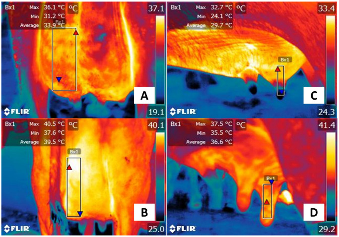
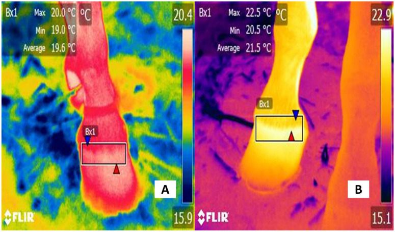
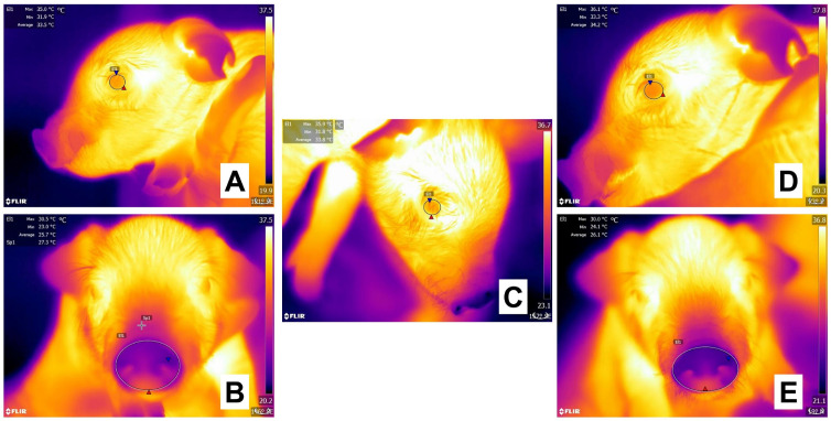
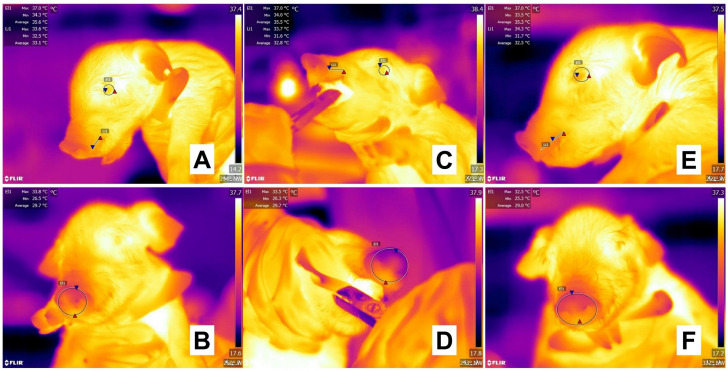
Pain assessment in domestic animals has gained importance in recent years due to the recognition of the physiological, behavioral, and endocrine consequences of acute pain on animal production, welfare, and animal model validity. Current approaches to identifying acute pain mainly rely on behavioral-based scales, quantifying pain-related biomarkers, and the use of devices monitoring sympathetic activity. Infrared thermography is an alternative that could be used to correlate the changes in the superficial temperature with other tools and thus be an additional or alternate acute pain assessment marker. Moreover, its non-invasiveness and the objective nature of its readout make it potentially very valuable. However, at the current time, it is not in widespread use as an assessment strategy. The present review discusses scientific evidence for infrared thermography as a tool to evaluate pain, limiting its use to monitor acute pain in pathological processes and invasive procedures, as well as its use for perioperative monitoring in domestic animals.

Keywords: analgesia; castration; inflammatory response; infrared thermography (IRT); invasive procedures; nociception; surgery; teeth clipping.


Conflict of interest statement

The authors declare no conflict of interest.

Figures

Figure 1
Figure 1
Nociceptive pathway during laminitis. The inflammatory process and tissue injury caused by laminitis trigger the first phase of nociception. (1) Transduction. The noxious stimulus is recognized and transformed into an electrical signal by peripheral nerves (Aδ and C fibers), known as nociceptors. In these free nerve endings, receptors such as ASIC3 or TRPV1, among others, are activated to create action potentials that will be transmitted to the DRG of the spinal cord. (2) Transmission. Through Aδ and C fibers, the noxious input is transmitted to the spinal cord, to synapse with second-order neurons in the gray matter of the structure. (3) Modulation. Once the signal reaches the spinal cord, spinal interneurons are responsible for projecting or modulating the signal by releasing inhibitory or excitatory neurotransmitters. (4) Projection. Through the spinothalamic tract, the electrical signal reaches superior centers in the brain, mainly the thalamus. (5) Perception. From the thalamus, third-order neurons project to the somatosensory cortex, where the conscious recognition of pain is developed. Due to the interaction of the thalamus with other regions such as the hypothalamus, pain activates sympathetic centers, which causes physiological, endocrine, and behavioral responses. The vasomotor response, occurring as a result of pain and inflammation, causes vasodilation in the injured site to promote immune cells invasion to the injury site, with consequent healing. The increase in blood flow in the region also increases the amount of radiated heat from the skin. This element is captured by thermal cameras, helping to identify an inflammatory process in animals. ASIC: acid-sensing ion channel; DRG: dorsal root ganglion; TRPV1: transient potential receptor vanilloid 1.
Figure 2
Figure 2
The local inflammatory response in the udder of a patient with mastitis. (1) The presence of a tissue injury or pathogens initiates inflammation by releasing local chemical compounds. (2) First-line mediators such as histamine, NO, ROS, and ions induce vasodilation to increase the permeability of the blood vessels and blood flow. (3) Leukocyte activation, emigration, and migration cause the presence and degranulation of neutrophils and other inflammatory cells into the injury site. (4) Release of pro-inflammatory substances such as prostaglandins, leukotrienes, cytokines, and neuropeptides, among others, exacerbates the inflammatory response to promote pathogens’ phagocytosis, and healing IL: interleukin; NK: neurokinin; NO: nitric oxide; SP: substance P; VIP: vasoactive intestinal polypeptide.
Figure 3
Figure 3
The systemic inflammatory response in pneumonia. Infection of the airway epithelium by pathogens leads to an inflammatory response where the mediastinal lymph node has a crucial role by participating in trafficking by DC, known as the take up of pathogens to the lymph node. Within the lymph node, antigen presentation takes place on naïve T cells to release effector T cells and induce the adaptative response of the organism. Degranulation of monocyte and polymorphic cells into the bloodstream causes a cytokine storm (SP, NK, IL), a reaction that promotes systemic inflammation and clinical signs. DC: dendritic cells; IL: interleukin; NK: neurokinin; SP: substance P.
Figure 4
Figure 4
Superficial thermal response associated with acute pain in a horse with colic. (A,C). Healthy female quarter-horse patient. The maximum temperature in the lacrimal caruncle (El1) was 36.8 °C (red triangle), and the minimum was 34.6 °C (blue triangle). In the caudal abdominal region (Bx1), the surface temperature presented a maximum temperature of 33.9 °C (red triangle) and a minimum of 29 °C (blue triangle). (B) Thoroughbred female equine with acute pain associated with colic. It is observed that the maximum (red triangle) and minimum (blue triangle) temperatures of the lacrimal caruncle (El1) decreased by 2.8 °C and 3.4 °C, respectively, compared to the healthy horse. (D) Contrary to the thermal response in the lacrimal caruncle, in the caudal abdominal region (Bx1) of the sick animal, the maximum surface temperature increased by 2.5 °C. The thermal images were taken by the authors of the present review. Radiometric images were obtained using a T1020 FLIR thermal camera. Image resolution 1024 × 768; up to 3.1 MP with UltraMax. FLIR Systems, Inc. Wilsonville, OR, USA.
Figure 5
Figure 5
Thermographic evaluation of the thermal response of the mammary gland in dairy cows and buffaloes. (A) Healthy udder of a Holstein bovine. The surface temperature of the caudal quarter (Bx1) of the udder shows a maximum temperature of 36.1 °C (red triangle) and a minimum value of 31.2 °C (blue triangle). (B) Udder of a Holstein cow with mastitis. The maximum and minimum temperature of the caudal quarter (Bx1) is 4.4 °C and 6.4 °C higher, respectively. (C) Healthy udder in a Murrah water buffalo. The surface temperature of the healthy lateral caudal quarter (Bx1) recorded a maximum temperature of 32.7 °C (red triangle) and a minimum of 24.1 °C (blue triangle). (D) Udder with mastitis in dairy buffalo. Compared with the thermal response of the healthy udder, the temperature of the caudal quarter (Bx1) of the udder is 4.8 °C higher at the maximum temperature (red triangle) and 11.4 °C of the minimum temperature (blue triangle) compared to the temperature of the healthy udder. Infectious agents such as Escherichia coli or Staphylococcus sp. can trigger the release of several pro-inflammatory cytokines that cause vasodilation and increase heat radiation associated with acute pain. The thermal images were taken by the authors of the present review. Radiometric images were obtained using a T1020 FLIR thermal camera. Image resolution 1024 × 768; up to 3.1 MP with UltraMax. FLIR Systems, Inc. Wilsonville, OR, USA.
Figure 6
Figure 6
Thermographic evaluation of hoof inflammation due to laminitis. (A) Healthy hoof. The hoof of a quarter-horse male with no reported pathologies. A maximum temperature of 20 °C (red triangle) and a minimum of 19 °C (blue triangle) can be observed on the surface of the coronet band (Bx1). (B) Hoof with laminitis. The thermal response of the coronet band (Bx1) of a female horse of the Azteca breed recorded increases by 2.5 °C and 1.5 °C at the maximum (red triangle) and minimum temperature (blue triangle). This may be due to the invasion of infectious agents during laminitis, which can trigger the release of interleukin-1, interleukin-10, histamine, and prostaglandin F2 alpha, causing an increase of heat in the inflamed area. The thermal images were taken by the authors of the present review.
Figure 7
Figure 7
Evaluation of the thermal response of 1-day-old piglets during tail docking with side-cutter pliers. (A,B) Before tail docking, the ocular surface (A, El1) of the piglet registered a maximum temperature (red triangle) of 35.0 °C, while the nasal window (B, El1) had a maximum temperature (red triangle) of 30.5 °C. (C). During the procedure, the ocular surface window increased its maximum temperature by 0.9 °C. (C,D) When comparing basal values with the surface temperatures taken immediately after tail docking, the maximum temperature of the piglet at the ocular window (D, El1) increased by 1.1 °C. At the same time, the nasal region (E, El1) decreased by 0.5 °C. This ambivalent reaction could be a result of the HPA axis increasing blood flow to important organs such as the eye and limiting it to a peripheral region such as the piglet’s nose. Sp1: default focal point of the software. The thermal images were taken by the authors of the present review. Radiometric images were obtained using a T1020 FLIR thermal camera. Image resolution 1024 × 768; up to 3.1 MP with UltraMax. FLIR Systems, Inc. Wilsonville, OR, USA.
Figure 8
Figure 8
Evaluation of the thermal response in White large x Landrace piglets during teeth clipping. (A,B) Before teeth clipping in 1-day-old piglets, the thermal windows of the ocular surface (A, El1), upper lip (Li1), and nose (B, El1) show a maximum temperature (red triangle) of 37.0 °C, 33.6 °C, and 33.8 °C, respectively. (C,D) During the procedure performed with clippers, the ocular surface’s maximum temperature (red triangle) (C, El1) maintained the same value as basal recordings. However, the maximum temperature of the upper lip (Li1) increased by 0.1 °C and in the nasal window (D, El1) decreased by 0.3 °C. (E,F) After the routine procedure, although the maximum temperature of the ocular surface (E, El1) did not change compared to basal values, a progressive increase was observed in the upper lip (Li1) by 0.7 °C. The drop in the maximum temperature (red triangle) of the nasal region (F, El1) by 1.3 °C shows the physiological response of animals when perceiving potential painful stimuli. The thermal images were taken by the authors of the present review. Radiometric images were obtained using a T1020 FLIR thermal camera. Image resolution 1024 × 768; up to 3.1 MP with UltraMax. FLIR Systems, Inc. Wilsonville, OR, USA.
Figure 9
Figure 9
Thermal response associated with acute pain in dogs undergoing surgery. (A) Before surgery. A 1-year-old bitch of the Chihuahua breed was subjected to ovariohysterectomy under analgesic management with meloxicam (0.1 mg/kg IV). The maximum temperature of the lacrimal caruncle (El1) had values of 36.6 °C (red triangle), while the minimum was 35.1 °C (blue triangle). (B) 1 h after surgery. A decrease in the temperature of the lacrimal caruncle is observed, with maximum (red triangle) and minimum (blue triangle) values of 35.3 °C and 32.6 °C, respectively. This represents a decrease of 1.3 °C and 2.5 °C, respectively. This could represent a sympathetic response to the effect of pain despite the use of analgesics. (C) 2 h after surgery. After administering tramadol as rescue analgesia, the maximum and minimum temperatures of the lacrimal caruncle (El1) (37.0 °C and 34.0 °C) were recorded above the basal values, due to the effect of the drug that reduces sympathetic tone. (D) 3 h post-surgery. Although it is observed that the maximum temperature of the lacrimal caruncle (El1) decreased by 0.6 °C (red triangle), the minimum increased by 0.3 °C (blue triangle), which could be associated with post-surgical stability. The thermal images were taken by the authors of the present review. Radiometric images were obtained using a T1020 FLIR thermal camera. Image resolution 1024 × 768; up to 3.1 MP with UltraMax. FLIR Systems, Inc. Wilsonville, OR, USA.
Figure 10
Figure 10
Mechanism of action and effect of analgesics in the nociceptive response and local inflammation. After tissue injury, the released pro-inflammatory mediators (e.g., NO, H+, histamine, 5-HT, among others) extend the local reaction to produce the five cardinal signs of inflammation. The administration of analgesic drugs, for example, NSAIDs, lessens this reaction by inhibiting the cyclooxygenase enzyme. In this way, reactions promoted by prostaglandin and prostacyclin, such as vasodilatation, pain, and local and systemic hyperthermia can be prevented. 5-HT: serotonin; ATP: adenosine triphosphate; BRK: bradykinin; H+: hydrogen; IL: interleukin; NGF: nerve growth factor; NO: nitric oxide; NSAIDs: non-steroidal anti-inflammatory drugs; PGE2: prostaglandin E2; TNFα: tumor necrosis factor alpha.
Figure 11
Figure 11
Thermal images in Wistar rats exposed to euthanasia with CO2. (A,B) Before the application of the euthanasia method, the ocular surface (El1), the auricular window (El2), and the base of the tail (Sp1) had a maximum temperature of 36.8 °C, 38.5 °C, and 27.6 °C, respectively. (C,D) During the euthanasia of the rodent with CO2 exposition inside a chamber, a general drop in the maximum temperature of the ocular surface (El1) and tail base (Sp1) by 0.9 °C and 1.5 °C was recorded. In contrast, an increase in the maximum temperature of the auricular region was reported (38.6 °C). (E,F) Within the first two minutes after exposition to CO2 euthanasia, all thermal windows showed a significant drop in temperature. When compared to basal values, the maximum temperature of the ocular region (El1) decreased by 2.5 °C, the auricular region (El2) by 2.8 °C, and the tail base (Sp1) by 2.2 °C. The progressive temperature drops in the rats, assessed by IRT, could help to determine the vasodilation effect of the euthanasia drug and could even be associated with pain when correlated with other evaluation tools. Maximal temperature is indicated with a red triangle and the minimal with a blue triangle. The thermal images were taken by the authors of the present review. Radiometric images were obtained using a T1020 FLIR thermal camera. Image resolution 1024 × 768; up to 3.1 MP with UltraMax. FLIR Systems, Inc. Wilsonville, OR, USA.
Figure 12
Figure 12
Recognition of inflammatory lesions in water buffaloes. (A,B) Thermal response due to lesion in the parietal region (red circle). An increase in the superficial temperature of the lacrimal caruncle (El1) can be associated with the lesion, with a maximum temperature of 40 °C (red triangle) and a minimum of 37.9 °C (blue triangle). (C,D) An ulcerative lesion on the elbow can be seen in the digital image. Through thermal imaging, the maximum temperature of the auricular region is 40 °C with a minimum of 33.6 °C. (E,F) The digital image shows a lesion in the shoulder region (red circle), whereas the radiometric image shows that the surface temperature of this region (El1) presented a maximum temperature of 38 °C (red triangle) and a minimum temperature of 31.8 °C (blue triangle), which would help to corroborate the presence of a lesion in the shoulder region as a result of the inflammatory process. The thermal images were taken by the authors of the present review.

Similar articles

Cited by

References

    1. Raja S.N., Carr D.B., Cohen M., Finnerup N.B., Flor H., Gibson S., Keefe F.J., Mogil J.S., Ringkamp M., Sluka K.A., et al. The revised International Association for the Study of Pain definition of pain: Concepts, challenges, and compromises. Pain. 2020;161:1976–1982. doi: 10.1097/j.pain.0000000000001939. - DOI - PMC - PubMed
    1. Mainau E., Manteca X. Pain and discomfort caused by parturition in cows and sows. Appl. Anim. Behav. Sci. 2011;135:241–251. doi: 10.1016/j.applanim.2011.10.020. - DOI
    1. Hernández-Avalos I., Mota-Rojas D., Mora-Medina P., Martínez-Burnes J., Casas Alvarado A., Verduzco-Mendoza A., Lezama-García K., Olmos-Hernandez A. Review of different methods used for clinical recognition and assessment of pain in dogs and cats. Int. J. Vet. Sci. Med. 2019;7:43–54. doi: 10.1080/23144599.2019.1680044. - DOI - PMC - PubMed
    1. Hugonnard M., Leblond A., Keroack S., Cadoré J., Troncy E. Attitudes and concerns of French veterinarians towards pain and analgesia in dogs and cats. Vet. Anaesth. Anal. 2004;31:154–163. doi: 10.1111/j.1467-2987.2004.00175.x. - DOI - PubMed
    1. Rekant S.I., Lyons M.A., Pacheco J.M., Arzt J., Rodriguez L.L. Veterinary applications of infrared thermography. Am. J. Vet. Res. 2016;77:98–107. doi: 10.2460/ajvr.77.1.98. - DOI - PubMed

LinkOut - more resources