. 2023 Jul 5;13(13):2213.
doi: 10.3390/ani13132213.

New Definition of Neoprotereunetes Fain et Camerik, Its Distribution and Description of the New Genus in Eupodidae (Acariformes: Prostigmata: Eupodoidea)

Affiliations

New Definition of Neoprotereunetes Fain et Camerik, Its Distribution and Description of the New Genus in Eupodidae (Acariformes: Prostigmata: Eupodoidea)

Ronald Laniecki et al. Animals (Basel). .

Erratum in

Abstract

The genus Neoprotereunetes Fain et Camerik, 1994 is revised and its definition is extended in order to incorporate some species of the invalid genus Protereunetes Berlese, 1923. The former type species Neoprotereunetes-Ereunetes lapidarius Oudemans, 1906 is redescribed and transferred to Filieupodes Jesionowska, 2010 (Cocceupodidae); Proterunetes boerneri is redescribed and designated the new type species. Two species groups are proposed to embrace Arctic and Antarctic species, respectively. Protereunetes paulinae Gless, 1972 is redescribed, whereas Protereunetes maudae Strandtmann, 1967 is redescribed and designated the type species of the new genus Antarcteupodes gen. nov. A key to the species of Neopretereunetes is provided.

Keywords: Acari; Protereunetes; biogeography; polar regions; taxonomy.


Conflict of interest statement

The authors declare no conflict of interest.

Figures
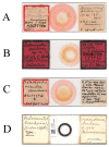
Figure 1
Figure 1
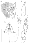
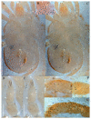
Microscopic slides. (A) Neoprotereunetes minutus (Strandtmann, 1967), holotype male; (B) Neoprotereunetes paulinae (Gless, 1972), holotype female; (C) Antarcteupodes maudae (Strandtmann, 1967), holotype female; (D) Filieupodes lapidarius (Oudemans, 1906), holotype female.
Figure 2
Figure 2
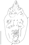
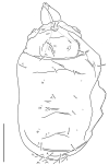
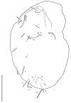
Neoprotereunetes boerneri (Thor, 1934), female. Body, dorsal view. Scale bar: 50 μm.
Figure 3
Figure 3
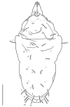
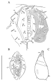
Neoprotereunetes boerneri (Thor, 1934), female. Body, ventral view. Scale bar: 50 μm.
Figure 4
Figure 4
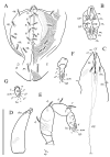
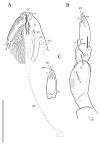
Neoprotereunetes boerneri (Thor, 1934), female. (A) Genital and anal region; (B) progenital chamber; (C) subcapitulum, ventral view; (D) right chelicera, lateral view; (E) left palp, lateral view; (F) tarsus of left palp, dorsal view; (G) tarsus of right palp, apical view. Scale bar: 50 μm.
Figure 5
Figure 5
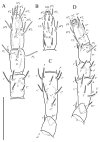
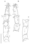
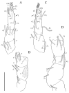
Neoprotereunetes boerneri (Thor, 1934), female. (A) Tarsus, tibia and genu of right leg I, dorsal view; (B) tarsus of right leg I, ventral view (apotele omitted); (C) femur and trochanter of right leg I, dorsal view; (D) right leg II, dorsolateral view. Asterisk denotes unpaired tarsal seta. Scale bar: 50 μm.
Figure 6
Figure 6
Neoprotereunetes boerneri (Thor, 1934), female. (A) Right leg III, dorsolateral view; (B) right leg IV, lateral view. Scale bar: 50 μm.
Figure 7
Figure 7
Neoprotereunetes boerneri (Thor, 1934), female. (A) Body, dorsal view; (B) naso; (C) body, ventral view; (D) tarsus and tibia of left palp, dorsal view; (E) tarsus and tibia of left palp, ventral view; (F) tarsus and tibia of right leg I, dorsal view; (G) tarsus of right leg I, ventral view; H—tarsus and tibia of right leg II, dorsolateral view. Asterisk denotes unpaired tarsal seta. Scale bar: (A,C) 100 μm; (B) 10 μm; (DH) 20 μm.
Figure 8
Figure 8
Neoprotereunetes paulinae (Gless, 1972), holotype female. Body, dorsal view. Scale bar: 100 μm.
Figure 9
Figure 9
Neoprotereunetes paulinae (Gless, 1972), holotype female. Body, ventral view. Scale bar: 100 μm.
Figure 10
Figure 10
Neoprotereunetes paulinae (Gless, 1972), holotype female. (A) genital region; (B) subcapitulum, ventral view; (C) left chelicera, lateral view; (D) left palp, dorsolateral view; (E) tarsus of right palp, dorsolateral view. Scale bar 50 μm.
Figure 11
Figure 11
Neoprotereunetes paulinae (Gless, 1972), holotype female. (A) Tarsus, tibia and genu of left leg I, dorsal view; (B) tarsus of left leg I, lateral view; (C) femur and trochanter of left leg I, dorsal view; (D) trochanter of right leg I, dorsal view; (E) left leg II, dorsolateral view. Scale bar: 50 μm.
Figure 12
Figure 12
Neoprotereunetes paulinae (Gless, 1972), holotype female. (A) Left leg III, dorsal view; (B) left leg IV, lateral view. Scale bar: 50 μm.
Figure 13
Figure 13
Neoprotereunetes paulinae (Gless, 1972), holotype female. (A) Body, dorsal and ventral view; (B) prodorsum; (C) genital region (D) tarsus and tibia of left palp, lateral view, antiaxial facesurface; (E) tarsus and tibia of left palp, lateral view, paraxial facesurface; (F) tarsus and tibia of right leg I, lateral view; (G) tarsus, tibia and genu of right leg II, ventrolateral view. Scale bar: (A) 100 μm; (BG) 20 μm.
Figure 14
Figure 14
Antarcteupodes maudae (Strandtmann, 1967), holotype female. Body, dorsal view. Scale bar: 100 μm.
Figure 15
Figure 15
Antarcteupodes maudae (Strandtmann, 1967), holotype female. Body, ventral view. Scale bar: 100 μm.
Figure 16
Figure 16
Antarcteupodes maudae (Strandtmann, 1967), holotype female. (A) Genital region; (B) progenital chamber; (C) left chelicera, dorsal view. Scale bar: 50 μm.
Figure 17
Figure 17
Antarcteupodes maudae (Strandtmann, 1967), holotype female. (A) Subcapitulum, ventral view; (B) left palp, dorsal view; (C) tarsus of right palp, dorsal view. Scale bar: 50 μm.
Figure 18
Figure 18
Antarcteupodes maudae (Strandtmann, 1967), holotype female. (A) Tarsus, tibia and genu of left leg I, dorsal view; (B) tarsus of left leg I, ventral view; (C) femur and trochanter of left leg I, dorsal view; (D) left leg II, dorsolateral view. Scale bar: 50 μm.
Figure 19
Figure 19
Antarcteupodes maudae (Strandtmann, 1967), holotype female. (A) Left leg III, dorsal view; (B) tarsus, tibia and genu of left leg IV, dorsolateral view; (C) telo-, basifemur and trochanter of left leg IV, dorsolateral view. Scale bar: 50 μm.
Figure 20
Figure 20
Antarcteupodes maudae (Strandtmann, 1967), holotype female. (A) Body, dorsal view; (B) naso; (C) body, ventral view; (D) tarsus and tibia of right palp, dorsal view; (E) tarsus and tibia of right palp, ventral view; (F) tarsus and tibia of right leg I, dorsolateral view; (G) tarsus and tibia of right leg II, dorsolateral view. Scale bar: (A,C) 100 μm; (B) 10 μm; (DG) 20 μm.
Figure 21
Figure 21
Filieupodes lapidarius (Oudemans, 1906), holotype female. Idiosoma, dorsolateral view. Scale bar: 100 μm.
Figure 22
Figure 22
Filieupodes lapidarius (Oudemans, 1906), holotype female. Body, ventral view. Scale bar: 100 μm.
Figure 23
Figure 23
Filieupodes lapidarius (Oudemans, 1906), holotype female. (A) Genital region; (B) subcapitulum, ventral view; (C) left chelicera, lateral view; (D) left palp, dorsal view. Scale bar: 50 μm.
Figure 24
Figure 24
Filieupodes lapidarius (Oudemans, 1906), holotype female. (A) Tarsus and tibia of right leg I, dorsolateral view; (B) genu, femur and trochanter of right leg I, dorsolateral view; (C) tarsus, tibia and genu of right leg II, dorsal view; (D) femur and trochanter of right leg II, dorsal view. Scale bar: 50 μm.
Figure 25
Figure 25
Filieupodes lapidarius (Oudemans, 1906), holotype female. (A) Tarsus, tibia and genu of right leg III, lateral view; (B) femur and trochanter of right leg III, lateral view; (C) tarsus and tibia of left leg IV, lateral view; (D) genu, femur and trochanter of left leg IV, lateral view. Scale bar: 50 μm.
Figure 26
Figure 26
Filieupodes lapidarius (Oudemans, 1906), holotype female. (A) Body, dorsal view; (B) prodorsum; (C) genital region; (D) tarsus and tibia of right palp, dorsal view; (E) tarsus and tibia of right palp, ventral view; (F) tarsus and tibia of right leg I, lateral view; (G) tarsus, tibia and genu of right leg II, dorsal view. Scale bar: (A) 100 μm; (BG) 20 μm.

References

    1. Krantz G.W., Walter D.E. A Manual of Acarology. 3rd ed. Texas Tech University Press; Lubbock, TX, USA: 2009. p. 807.
    1. Szudarek-Trepto N., Kaźmierski A., Dabert M., Dabert J. Molecular phylogeny of Eupodidae reveals that the family Cocceupodidae (Actinotrichida; Eupodoidea) and its genus Filieupodes are valid taxa. Exp. Appl. Acarol. 2020;80:43–57. doi: 10.1007/s10493-019-00455-4. - DOI - PubMed
    1. Baker A.S., Lindquist E.E. Aethosolenia laselvensis gen. n., sp. n., a new eupodoid mite from Costa Rica (Acari: Prostigmata) [(accessed on 1 November 2022)];Syst. Appl. Acarol. Spec. Publ. 2002 11:1–11. doi: 10.11158/saasp.11.1.1. Available online: https://www.nhm.ac.uk/hosted-sites/acarology/saas/saasp/2002/saasp11.pdf. - DOI
    1. Jesionowska K. Cocceupodidae, a new family of eupodoid mites, with description of a new genus and two new species from Poland. Part I. (Acari: Prostigmata: Eupodoidea) Genus. 2010;21:637–658.
    1. Gless E.E. Life cycle studies of some Antarctic mites and description of a new species, Protereunetes paulinae (Acari: Eupodidae) Antarct. Res. Ser. Wash. 1972;20:289–306.

LinkOut - more resources