. 2023 Jun 29;15(13):3413.
doi: 10.3390/cancers15133413.

Externally Applied Electromagnetic Fields and Hyperthermia Irreversibly Damage Cancer Cells

Affiliations

Externally Applied Electromagnetic Fields and Hyperthermia Irreversibly Damage Cancer Cells

Elena Obrador et al. Cancers (Basel). .

Abstract

At present, the applications and efficacy of non-ionizing radiations (NIR) in oncotherapy are limited. In terms of potential combinations, the use of biocompatible magnetic nanoparticles as heat mediators has been extensively investigated. Nevertheless, developing more efficient heat nanomediators that may exhibit high specific absorption rates is still an unsolved problem. Our aim was to investigate if externally applied magnetic fields and a heat-inducing NIR affect tumor cell viability. To this end, under in vitro conditions, different human cancer cells (A2058 melanoma, AsPC1 pancreas carcinoma, MDA-MB-231 breast carcinoma) were treated with the combination of electromagnetic fields (EMFs, using solenoids) and hyperthermia (HT, using a thermostated bath). The effect of NIR was also studied in combination with standard chemotherapy and targeted therapy. An experimental device combining EMFs and high-intensity focused ultrasounds (HIFU)-induced HT was tested in vivo. EMFs (25 µT, 4 h) or HT (52 °C, 40 min) showed a limited effect on cancer cell viability in vitro. However, their combination decreased viability to approximately 16%, 50%, and 21% of control values in A2058, AsPC1, and MDA-MB-231 cells, respectively. Increased lysosomal permeability, release of cathepsins into the cytosol, and mitochondria-dependent activation of cell death are the underlying mechanisms. Cancer cells could be completely eliminated by combining EMFs, HT, and standard chemotherapy or EMFs, HT, and anti-Hsp70-targeted therapy. As a proof of concept, in vivo experiments performed in AsPC1 xenografts showed that a combination of EMFs, HIFU-induced HT, standard chemotherapy, and a lysosomal permeabilizer induces a complete cancer regression.

Keywords: cancer cell death; cancer therapy; electromagnetic fields; hyperthermia; non-ionizing radiations.


Conflict of interest statement

The authors declare that no competing interest or personal relationship have influenced the work reported in this paper. R. López-Blanch M. Oriol-Caballo and M.P. Moreno-Murciano receive salary support from ScientiaBiotech.

Figures

Figure 1
Figure 1
Effect of EMFs and HT on cancer cell viability. (A) Effect of EMFs. Cancer cells were seeded and 24 h later exposed to EMFs (100–500 kHz × 1–5 h). Control values (0 kHz) were 1.14 ± 0.03 A2058, 1.13 ± 0.04 AsPC1, and 1.20 ± 0.05 MDA-MB-231 (×106) viable cells (n = 5 in all cases). * p < 0.05 comparing all conditions versus controls (0 kHz) (n = 5 t-test). (B) Effect of HT. Cancer cells were seeded and 24 h later exposed to HT (42–52 °C × 20–40 min). * p < 0.05 ** p < 0.01 comparing all conditions versus controls (37 °C) ++ p < 0.01 comparing 40 min versus 20 min (n = 5 t test). (C) Effect of EMFS and HT. Cancer cells were seeded and 24 h later exposed to EMFs (100 kHz × 4 h) and HT (52 °C × 40 min from min 120 to min 160 of the 4 h period where cells were constantly exposed to the EMFs). The surviving cells were cultured for 24 additional hours without further exposure to EMFs and HT. A two-way analysis of variance (ANOVA) was used to make comparisons among the different groups after 4 h of treatment with EMFs + HT and 24 h after. Different letters indicate differences p < 0.05 (n = 5).
Figure 2
Figure 2
Effect of EMFs and HT on cell cycle distribution and the type of death in cancer cells. (A) Flow cytometry analysis of the cell cycle distribution after exposure to EMFs and HT as in Figure 1C (n = 5). No statistically significant differences were found when comparing treatment with EMFs + HT and controls. (B) Flow cytometry analysis of EMFs and HT-induced apoptosis and necrosis (treatment as in Figure 1C). A two-way analysis of variance (ANOVA) was used to make comparisons among control cells treated with EMFs + HT and the different cell subpopulations in both groups. Different letters indicate differences p < 0.05 (n = 5).
Figure 2
Figure 2
Effect of EMFs and HT on cell cycle distribution and the type of death in cancer cells. (A) Flow cytometry analysis of the cell cycle distribution after exposure to EMFs and HT as in Figure 1C (n = 5). No statistically significant differences were found when comparing treatment with EMFs + HT and controls. (B) Flow cytometry analysis of EMFs and HT-induced apoptosis and necrosis (treatment as in Figure 1C). A two-way analysis of variance (ANOVA) was used to make comparisons among control cells treated with EMFs + HT and the different cell subpopulations in both groups. Different letters indicate differences p < 0.05 (n = 5).
Figure 3
Figure 3
Effect of EMFs and HT on the activation of apoptotic death in cancer cells. (A) Inverted microscope images (magnification ×10) of cancer cells treated with EMFs and HT (as in Figure 1C) showing the drastic morphological changes associated with the loss of viability. (B) Cell death analysis after the 4 h protocol (Figure 1C) based on Hoechst 33342 and propidium iodide (PI) staining and the TUNEL labeling assay (see under Methods). Cell viability in control flasks was > 98% in all cases. A one-way analysis of variance (ANOVA) was used to make comparisons among cell subsets. Different letters indicate statistical differences p < 0.05 (n = 5). (C) Western blots for detection of cytochrome C and AIF in the cytosolic fraction (all performed right after the 4 h protocol Figure 1C). Densitometric analysis (a.u. arbitrary units) represents the mean values ± SD for 5 different experiments per cell line [* p < 0.01 comparing cells treated with EMFs and HT (4 h as in Figure 1C) versus untreated controls t-test]. The original western blot figures could be found in Figure S3.
Figure 4
Figure 4
Effect of EMFs and HT on HSP70 and HSP110 and lysosomal permeability. (A) Protein levels (western blots) of Hsp70 and Hsp110 were measured in cancer cells after exposure to EMFs and HT (4 h protocol as in Figure 1C) and 24 h after exposure. Densitometric analysis (a.u. arbitrary units) represents the mean values ± SD for four different experiments per cell line and time point. A one-way analysis of variance (ANOVA) was used to make comparisons among the different experimental times. Different letters indicate statistical differences p < 0.05. (B) Cysteine and aspartate cathepsin activities in the cytosolic fraction were measured after exposure to EMFs and HT (4 h protocol as in Figure 1C) (n = 5 * p < 0.01 comparing EMFs and HT-treated cells versus untreated controls). (C) Lysosome staining (LysoTracker) was performed in the cancer cells (representative images) after the 4 h protocol (as in Figure 1C), showing EMFs and HT-induced diffusion of the lysosomal marker into the cytosol. The original western blot figures could be found in Figure S3.
Figure 5
Figure 5
Effect of EMFs HT and chemotherapy on cancer cell viability. Cancer cells were seeded 24 h before starting the treatments. Cells were treated with EMFs and HT (4 h), as in Figure 1C. Paclitaxel (PAC 1 μM) was present in the cultured medium during the 4 h protocol (Figure 1C). Gemcitabine (GEM 25 μM) was present in the cultured medium during the last 30 min of the 4 h protocol. At the end of the 4 h treatment period, the culture medium was changed, and the cells were kept in culture for 24 additional hours. A two-way analysis of variance (ANOVA) was used to make comparisons among the different groups after 4 h of treatment with EMFs + HT and 24 h after. Different letters indicate statistical differences p < 0.05 (n = 5).
Figure 6
Figure 6
Effect of EMFs HT and a natural lysosomal membrane permeabilizer or a targeted anti-Hsp70 therapy on cancer cells viability. Cells were treated with EMFs and HT, as in Figure 1C. Pterostilbene (PT 20 μM) or apoptozole (Az 4–5 μM depending on the IC50 values described in Section 3) were added to the cultured medium right before (PT) or 12 h before starting the 4 h protocol (Az) (as in Figure 1C). A one-way analysis of variance (ANOVA) was used to make comparisons among the different experimental groups. Different letters indicate statistical differences p < 0.05 (n = 5).
Figure 7
Figure 7
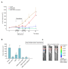
Effect of EMFs + HIFU-induced HT, gemcitabine and/or PT on the growth of AsPC1 pancreas carcinoma. Cancer cells were inoculated subcutaneously on day 0, and mice were treated with EMFs and HIFU as described under Materials and Methods. (A) EMFs and HIFU were applied once per day per three consecutive days (Monday to Wednesday) for two consecutive weeks starting on day 14 after tumor inoculation. Gemcitabine (50 mg/kg) was administered twice on days 14 and 21. A one-way analysis of variance (ANOVA) was used to make comparisons among the different experimental groups at each time point. Different letters indicate statistical differences p < 0.05 (n = 15 mice per experimental group). (B) A disodium salt of PT phosphate (Chromadex Inc. Los Angeles CA) (100 mg of PT/kg) was administered i.p. (one dose 30 min before starting each irradiation session with EMFs and HT). A one-way analysis of variance (ANOVA) was used to make comparisons among the different experimental groups. Different letters indicate statistical differences p < 0.05 (n = 12 mice per experimental group). (C) Representative images of mice inoculated with AsPC1/Luciferase Stable Cells and treated with EMFs HT and gemcitabine (GEM) or EMFs HT gemcitabine and PT.

References

    1. Saliev T., Begimbetova D., Masoud A.-R., Matkarimov B. Biological Effects of Non-Ionizing Electromagnetic Fields: Two Sides of a Coin. Prog. Biophys. Mol. Biol. 2019;141:25–36. doi: 10.1016/j.pbiomolbio.2018.07.009. - DOI - PubMed
    1. Wenger C., Miranda P.C., Salvador R., Thielscher A., Bomzon Z., Giladi M., Mrugala M.M., Korshoej A.R. A Review on Tumor-Treating Fields (TTFields): Clinical Implications Inferred from Computational Modeling. IEEE Rev. Biomed. Eng. 2018;11:195–207. doi: 10.1109/RBME.2017.2765282. - DOI - PubMed
    1. Chang F., Minc N. Electrochemical Control of Cell and Tissue Polarity. Annu. Rev. Cell Dev. Biol. 2014;30:317–336. doi: 10.1146/annurev-cellbio-100913-013357. - DOI - PubMed
    1. Krenacs T., Meggyeshazi N., Forika G., Kiss E., Hamar P., Szekely T., Vancsik T. Modulated Electro-Hyperthermia-Induced Tumor Damage Mechanisms Revealed in Cancer Models. Int. J. Mol. Sci. 2020;21:6270. doi: 10.3390/ijms21176270. - DOI - PMC - PubMed
    1. Arvind R., Chandana S.R., Borad M.J., Pennington D., Mody K., Babiker H. Tumor-Treating Fields: A Fourth Modality in Cancer Treatment, New Practice Updates. Crit. Rev. Oncol. Hematol. 2021;168:103535. doi: 10.1016/j.critrevonc.2021.103535. - DOI - PubMed

Grants and funding