. 2023 Jun 30;15(13):3432.
doi: 10.3390/cancers15133432.

Vitamin D Receptor Antagonist MeTC7 Inhibits PD-L1

Affiliations

Vitamin D Receptor Antagonist MeTC7 Inhibits PD-L1

Negar Khazan et al. Cancers (Basel). .

Abstract

Small-molecule inhibitors of PD-L1 are postulated to control immune evasion in tumors similar to antibodies that target the PD-L1/PD-1 immune checkpoint axis. However, the identity of targetable PD-L1 inducers is required to develop small-molecule PD-L1 inhibitors. In this study, using chromatin immunoprecipitation (ChIP) assay and siRNA, we demonstrate that vitamin D/VDR regulates PD-L1 expression in acute myeloid leukemia (AML) and myelodysplastic syndrome (MDS) cells. We have examined whether a VDR antagonist, MeTC7, can inhibit PD-L1. To ensure that MeTC7 inhibits VDR/PD-L1 without off-target effects, we examined competitive inhibition of VDR by MeTC7, utilizing ligand-dependent dimerization of VDR-RXR, RXR-RXR, and VDR-coactivators in a mammalian 2-hybrid (M2H) assay. MeTC7 inhibits VDR selectively, suppresses PD-L1 expression sparing PD-L2, and inhibits the cell viability, clonogenicity, and xenograft growth of AML cells. MeTC7 blocks AML/mesenchymal stem cells (MSCs) adhesion and increases the efferocytotic efficiency of THP-1 AML cells. Additionally, utilizing a syngeneic colorectal cancer model in which VDR/PD-L1 co-upregulation occurs in vivo under radiation therapy (RT), MeTC7 inhibits PD-L1 and enhances intra-tumoral CD8+T cells expressing lymphoid activation antigen-CD69. Taken together, MeTC7 is a promising small-molecule inhibitor of PD-L1 with clinical potential.

Keywords: AML; M2H assay; MDS; PD-L1; VDR; efferocytosis; small molecule; vitamin D.


Conflict of interest statement

R.K.S. and R.G.M. are listed as the co-inventors on U.S. (11,034,719), EU (3128996), and Canadian (2,982,270) patents assigned to the University of Rochester. The rest of the authors do not carry a conflict of interest.

Figures
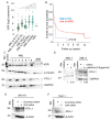
Figure 1
Figure 1
VDR is upregulated in AML subtypes and shows poor prognostication and upregulation of PD-L1 upon calcitriol stimulation. * p < 0.05; ** p < 0.01; (A) The VDR mRNA expression in AML patients and survival probability were analyzed using R2-Genomics Analysis and Visualization Platform (Bohlander-422-MAS5.0_u133a). (B) Overall survival in AML patients based on VDR expression (high: n = 8; low: n = 409) showed decreased survival. (C) Immunoblotting of VDR and PD-L1 in cells from randomly selected primary (n = 5) and relapsed (n = 2) AML patients, compared to normal bone marrow (nBM) (n = 3). (D) Immunoblotting of PD-L1 in AML-1 and AML-2 cells with calcitriol (400 nM) stimulation for 48 h. The densitometry analysis is shown below the bands. Immunoblotting of PD-L1 in MV-411 cells (E) and THP-1 cells (F) expressing scrambled siRNA or VDR siRNA. The uncropped blots are shown in Figure S3.
Figure 2
Figure 2
MeTC7 is a potent VDR antagonist. (A) Chemical structure of MeTC7. (B) MeTC7 demonstrates selective competitive inhibition. Heterodimerization of RXR-VDR in response to 1,25D and/or MeTC7, and homodimerization of RXR-RXR in response to bexarotene and/or MeTC7 in a mammalian 2-hybrid system (M2H). (C) VDR binding to receptor co-activator SRC1 in an M2H assay. (D) VDR binding to receptor co-activator DRIP205 in an M2H assay. (E) Selective competitive inhibition of VDR agonist 3-keto lithocholate by MeTC7. Results are plotted as ligand-mediated RXR-VDR heterodimerization (or RXR-RXR homodimerization) compared to the positive control (1,25D or bexarotene) set to 100%. The negative control is DMSO. Error bars represent standard deviations (* p < 0.05 versus 1,25D control). The data are a compilation of between six and eight independent assays, with each treatment group dosed in quadruplicate for each independent assay. The transcriptional activation of the reporter gene was measured in comparison to the reference compound 1,25D or bexarotene. Error bars indicate the standard deviation of the replicate experiments. Different shadings of blue in panels B-E represent the VDR-containing M2H systems, while different shadings of green represent the RXR-only M2H. White fill represents the DMSO negative (vehicle) control.
Figure 3
Figure 3
MeTC7 selectively inhibits PD-L1 expression in AML cells: MeTC7 treatment reduced PD-L1 expression in AML-1 primary cells (A), MV-411 (B), and THP-1 (C) cells during 48 h of indicated dose treatment. MeTC7 treatment at the indicated doses did not alter the expression of PD-L2 in THP-1 and MV-411 cells (D,E). The uncropped blots are shown in Figure S5.
Figure 4
Figure 4
MeTC7 inhibited the proliferation of AML cells dose dependently. MV411, THP1, U937, and MOLM13 cell lines (Aleft),five randomly selected primary human AML cells (Amiddle) or three relapsed AML cells (Aright) were seeded and treated with DMSO or MeTC7 in complete RPMI-1640 medium for 40 h. MTS (Promega, Celltiter96Aqueous one solution, #G3580) (10 µL) was added to each well and to those without cells (blank) and incubated for an additional 4–6 h. The optical density of the wells was recorded using the BioRad iMark microplate reader at 490 nM wavelength. The percentage viability of MeTC7-treated cells was calculated relative to DMSO-treated cells. Data as mean ± SEM are shown. (B) Table: Cytogenetic characteristics of five human primary AML cells used in Figure 5B are shown. MeTC7 treatment inhibited colonies formed by THP-1 (Cleft) and U937 (Cright) cells. THP1 and U937 cells were added to MethoCult H4435 Enriched (Stem Cell Technologies, #: 04435) to achieve a concentration of 2000 cells/mL. DMSO (<0.2%) or MeTC7 (250 and 500 nM) was added. Each condition was performed in triplicate. The plates were incubated at 37 °C with 5% CO2 in a humidified incubator. Colonies were counted on day 7 (U937 cells) and day 14 (THP1 cells). One-way ANOVA and Tukey’s multiple comparison tests of the number of colonies formed in control versus treatment groups were performed using GraphPrism-8. * p < 0.05; ** p < 0.005, *** p < 0.0005, **** p < 0.00005. (D) Representative light contrast images of the U937 cell colonies treated with vehicle or MeTC7 (250 and 500 nM) on day 10 of the experiment: (Cright) growing into MethoCult H4435 enriched (Stem Cell Technologies, Canada, # 04435) are shown. (E) MeTC7 treatment reduced the colony units formed by a primary AML cell in MethoCult H4435 enriched dose dependently. Ara-C was used as a positive control. Details of the experimental procedures are described in the Supplementary Information section.
Figure 5
Figure 5
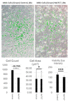
Effect of MeTC7 on the attachment of primary AML cells with MSCs. MSCs were co-cultured with pre-stained CellTracker Green for 24 h. Control (DMSO) and MeTC7 (300 nM) treatment occurred by the addition of media-containing treatment into an equal volume of co-culture-containing media. At 24 and 48 h after treatment, cells were imaged using an Olympus CKX-41 inverted microscope equipped with epifluorescence illumination and the U-FF fluorescence filter kit (excitation 467–498 nm). Image acquisition used Olympus cellSens imaging software version 2.3. Background subtracted fluorescence images were merged with brightfield images using Adobe Photoshop, release 23.2.2. Annotation for * p < 0.05 and *** p < 0.0005. For area quantification, ImageJ (Fiji) software (ImageJ 1.53t, Java 1.8.0_322 (64-bit) was used. Randomly selected AML cells (n = 400) from each population were manually traced. Statistical analyses were performed using n = 400 respective cell populations. Area of traced cell calculated by software was calibrated to image parameters. Cell viability (dye intensity) was determined semi-quantitatively utilizing the color expression via the reciprocal intensity of images using ImageJ (ImageJ 1.53t, Java 1.8.0_322 (64-bit).
Figure 6
Figure 6
MeTC7 enhances the phagocytotic activity of THP1 cells. THP1 cells were treated with MeTC7 (340 nM) or vehicle for 48 h. Neutrophils (PMNs), isolated from human peripheral blood, were incubated at −80 °C in FBS+ 10% DMSO for 24 h to induce apoptosis. Apoptotic neutrophils were washed with PBS and fluorescently labeled with 2 μM PKH26GL. End-stage neutrophils were provided in excess (10:1) to plated THP1 cells for 3 h. Cells were imaged and collected for flow cytometry analysis. All samples were run on an LSRII flow cytometer using FSC, SSC, 355 nm (DAPI), and 535 nm (PKH26). Analysis was performed using FlowJo version 10.7.1. Annotation for ns: not significant, **** p < 0.0001. Images were taken at 20×. Scale bar = 50 µM.
Figure 7
Figure 7
MeTC7 reduces PD-L1 expression in an RT-inducible PD-L1overexpression syngeneic model. (A): Schema of determination of activity against PD-L1 activated by RT in an orthotopic model of MC38 colorectal cancer in BL7 mice. MeTC7, in combination with RT-abrogated RT-induced PD-L1 activation in CD45 tumor cells post-RT (B); (C), increased CD8+T-cell infiltration in tumors significantly compared to vehicle or RT + vehicle. (D,E): MeTC7 increased the %of cells expressing CD69 (D) and PD-1 (E) compared to vehicle and vehicle + RT. The details of the antibodies used are listed in Supplementary Information S1. Annotation for * p < 0.05.
Figure 8
Figure 8
Proposed signaling effects of MeTC7 against VDR/PD-L1 in cancer cells.

References

    1. Dong H., Zhu G., Tamada K., Chen L. B7-H1, a third member of the B7 family, co-stimulates T-cell proliferation and interleukin-10 secretion. Nat. Med. 1999;5:1365–1369. doi: 10.1038/70932. - DOI - PubMed
    1. Dong H., Strome S.E., Salomao D.R., Tamura H., Hirano F., Flies D.B., Roche P.C., Lu J., Zhu G., Tamada K., et al. Tumor-associated B7-H1 promotes T-cell apoptosis: A potential mechanism of immune evasion. Nat. Med. 2002;8:793–800. doi: 10.1038/nm730. - DOI - PubMed
    1. Vandenborre K., Van Gool S.W., Kasran A., Ceuppens J.L., Boogaerts M.A., Vandenberghe P. Interaction of CTLA-4 (CD152) with CD80 or CD86 inhibits human T-cell activation. Immunology. 1999;98:413–421. doi: 10.1046/j.1365-2567.1999.00888.x. - DOI - PMC - PubMed
    1. ElTanbouly M.A., Schaafsma E., Noelle R.J., Lines J.L. VISTA: Coming of age as a multi-lineage immune checkpoint. Clin. Exp. Immunol. 2020;200:120–130. doi: 10.1111/cei.13415. - DOI - PMC - PubMed
    1. Lamberti G., Sisi M., Andrini E., Palladini A., Giunchi F., Lollini P.L., Ardizzoni A., Gelsomino F. The mechanisms of PD-L1 regulation in non-small-cell lung cancer (NSCLC): Which are the involved players? Cancers. 2020;12:3129. doi: 10.3390/cancers12113129. - DOI - PMC - PubMed