

Prediction of 2-[18F]FDG PET-CT SUVmax for Adrenal Mass Characterization: A CT Radiomics Feasibility Study
- PMID: 37444549
- PMCID: PMC10340369
- DOI: 10.3390/cancers15133439
Prediction of 2-[18F]FDG PET-CT SUVmax for Adrenal Mass Characterization: A CT Radiomics Feasibility Study
Abstract
Background: Indeterminate adrenal masses (AM) pose a diagnostic challenge, and 2-[18F]FDG PET-CT serves as a problem-solving tool. Aim of this study was to investigate whether CT radiomics features could be used to predict the 2-[18F]FDG SUVmax of AM.
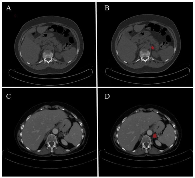
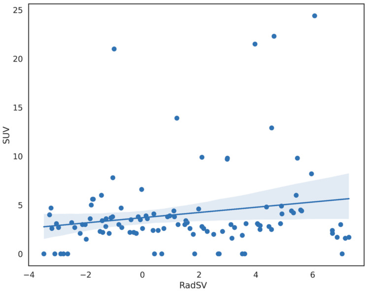
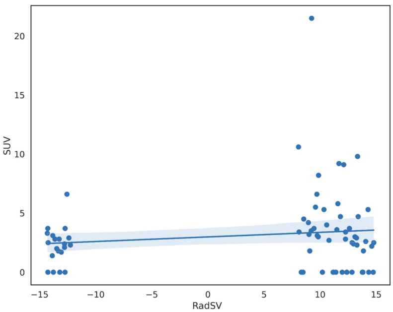
Methods: Patients with AM on 2-[18F]FDG PET-CT scan were grouped based on iodine contrast injection as CT contrast-enhanced (CE) or CT unenhanced (NCE). Two-dimensional segmentations of AM were manually obtained by multiple operators on CT images. Image resampling and discretization (bin number = 16) were performed. 919 features were calculated using PyRadiomics. After scaling, unstable, redundant, and low variance features were discarded. Using linear regression and the Uniform Manifold Approximation and Projection technique, a CT radiomics synthetic value (RadSV) was obtained. The correlation between CT RadSV and 2-[18F]FDG SUVmax was assessed with Pearson test.
Results: A total of 725 patients underwent PET-CT from April 2020 to April 2021. In 150 (21%) patients, a total of 179 AM (29 bilateral) were detected. Group CE consisted of 84 patients with 108 AM (size = 18.1 ± 4.9 mm) and Group NCE of 66 patients with 71 AM (size = 18.5 ± 3.8 mm). In both groups, 39 features were selected. No statisticallyf significant correlation between CT RadSV and 2-[18F]FDG SUVmax was found (Group CE, r = 0.18 and p = 0.058; Group NCE, r = 0.13 and p = 0.27).
Conclusions: It might not be feasible to predict 2-[18F]FDG SUVmax of AM using CT RadSV. Its role as a problem-solving tool for indeterminate AM remains fundamental.
Keywords: PET-CT; SUVmax; adrenal glands; neoplasms; radiomics.
Conflict of interest statement
The authors declare no conflict of interest.
Figures
References
LinkOut - more resources
Full Text Sources
Miscellaneous
