. 2023 Jun 26;11(13):1862.
doi: 10.3390/healthcare11131862.

The Effectiveness of Compression Garments for Reducing Pain in Non-Vascular Ehlers-Danlos Syndromes: A Prospective Observational Cohort Study

Affiliations

The Effectiveness of Compression Garments for Reducing Pain in Non-Vascular Ehlers-Danlos Syndromes: A Prospective Observational Cohort Study

Karelle Benistan et al. Healthcare (Basel). .

Abstract

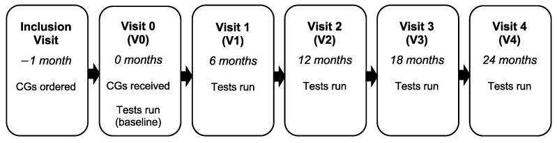
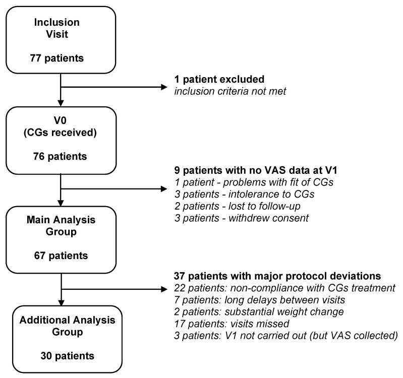
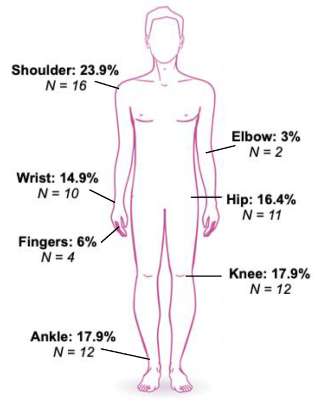
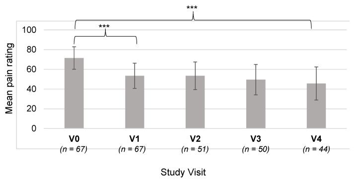
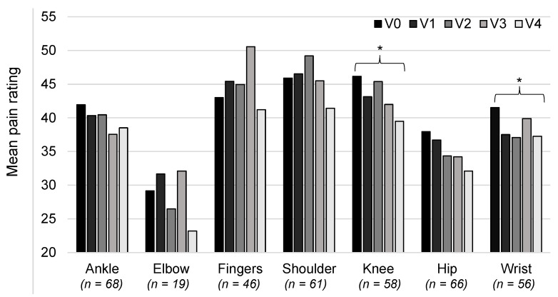
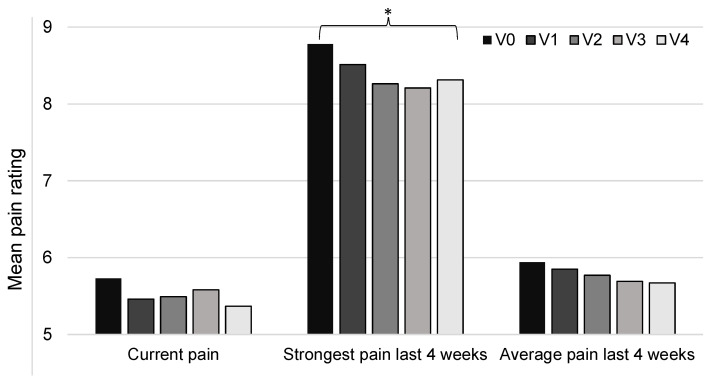
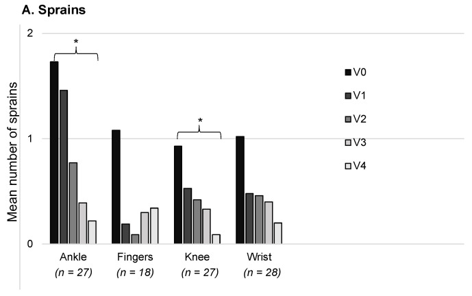
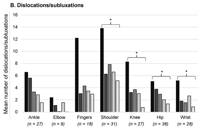
Patients with Ehlers-Danlos Syndrome (EDS) frequently suffer from severe chronic pain. We carried out an observational cohort study to assess the effectiveness of compression garments (CGs) for reducing this pain. Patients with non-vascular EDS were given custom-made Cerecare® CGs during a visit to a specialist clinic (visit V0). They were followed up over 2 years with visits every 6 months (V1-V4). At each visit, pain was assessed for the joints treated with CGs using a visual analogue scale (VAS; 0-100 mm). Additional measures were obtained to assess neuropathic pain (painDETECT questionnaire), proprioception/balance (Berg Balance Scale), and functional independence, amongst others. Data were analyzed for 67 patients with EDS (hypermobile: 91%; classical: 6%; kyphoscoliotic: 3%). For the most painful joint, the mean VAS rating was 71.5 ± 22.8 mm at V0; this decreased to 53.5 ± 25.5 mm at V1 and 45.7 ± 29 mm at V4 (t-tests: p < 0.0001). From V0 to V4, improvements were also seen for pain at the other joints, neuropathic pain, functional independence, proprioception/balance, and the incidence of sprains and dislocations/subluxations, although not all comparisons were statistically significant (p < 0.05 level). These results indicate that CGs may effectively reduce the pain and joint instability in non-vascular EDS patients.

Keywords: Ehlers-Danlos syndromes; compression garments; dislocation; joint hypermobility; joint instability; orthoses; pain therapy; proprioception; quality of life; sprains.


Conflict of interest statement

The authors declare no conflict of interest. The funders had no role in the design of the study; in the collection, analyses, or interpretation of data; in the writing of the manuscript; or in the decision to publish the results.

Figures

Figure 1
Figure 1
The patients’ visits to the clinic.
Figure 2
Figure 2
Participant flow diagram. Abbreviations: CGs: compression garments; VAS: visual analogue scale for pain.
Figure 3
Figure 3
The percentage of patients prescribed each compression garment in the main analysis group (n = 67). Means are shown for items that could be for the left or right side (e.g., elbow sleeves), and for gloves and mittens with or without fingers. The garment supplements are indicated by a plus sign. Abbreviations: L: left; R: right.
Figure 4
Figure 4
The length of time the compression garments were worn each day. The percentage of patients who wore the garments for <6, 6–12, 12–24, or 24 h is shown, as assessed at visits V1 to V4. Data are shown for shorts and vests.
Figure 5
Figure 5
The most painful joint prior to wearing the compression garments. The number of patients who reported each joint to be the most painful is shown, along with the percentages.
Figure 6
Figure 6
Mean pain ratings over the course of the study for the most painful joint. Pain was assessed using a visual analogue scale from 0–100 mm. The number of patients for each time point is shown along the x axis. *** p < 0.0001; error bars: standard deviation.
Figure 7
Figure 7
Mean pain ratings over the course of the study for each joint treated with compression garments. Pain was assessed using a visual analogue scale (0–100 mm). The number of patients with data at V4 are shown along the x axis; note that one patient may be counted twice due to data for the right and left sides. * p < 0.05.
Figure 8
Figure 8
Measures of pain from the painDETECT questionnaire. Pain was rated on a scale of 0–10. There were data for 67 patients at V0 and V1, 57 at V2, 52 at V3, and 49 at V4. * p < 0.05.
Figure 9
Figure 9
Mean scores for neuropathic pain at the most painful area. Patients responded to items on a scale, and these were converted into a score from 0 to 5. There were data for up to 67 patients at V0 with decreasing numbers down to 49 at V4. * p < 0.05.
Figure 10
Figure 10
Mean number of sprains and dislocations/subluxations over a 30-day period. The number of patients with data for their joints treated with CG for the duration of study is shown along the x axis. Note that one patient may be counted twice due to data for the right and left sides. * p < 0.05.
Figure 10
Figure 10
Mean number of sprains and dislocations/subluxations over a 30-day period. The number of patients with data for their joints treated with CG for the duration of study is shown along the x axis. Note that one patient may be counted twice due to data for the right and left sides. * p < 0.05.
Figure 11
Figure 11
Percentage of patients who reported improvement, worsening, or stability of wounds, scars, bruises, and stretch marks at V1. For each skin condition, the central bar (dark gray) indicates stability, the bar to the right indicates an improvement, and the bar to the left indicates worsening. Note that the percentages do not total 100% because some patients responded that they did not know (not plotted).

References

    1. Malfait F., Castori M., Francomano C.A., Giunta C., Kosho T., Byers P.H. The Ehlers-Danlos Syndromes. Nat. Rev. Dis. Prim. 2020;6:64. doi: 10.1038/s41572-020-0194-9. - DOI - PubMed
    1. Tinkle B., Castori M., Berglund B., Cohen H., Grahame R., Kazkaz H., Levy H. Hypermobile Ehlers-Danlos Syndrome (a.k.a. Ehlers-Danlos Syndrome Type III and Ehlers-Danlos Syndrome Hypermobility Type): Clinical Description and Natural History. Am. J. Med. Genet. Part C Semin. Med. Genet. 2017;175:48–69. doi: 10.1002/ajmg.c.31538. - DOI - PubMed
    1. Malfait F., Francomano C., Byers P., Belmont J., Berglund B., Black J., Bloom L., Bowen J.M., Brady A.F., Burrows N.P., et al. The 2017 International Classification of the Ehlers-Danlos Syndromes. Am. J. Med. Genet. Part C Semin. Med. Genet. 2017;175:8–26. doi: 10.1002/ajmg.c.31552. - DOI - PubMed
    1. Bénistan K., Martinez V. Pain in Hypermobile Ehlers-Danlos Syndrome: New Insights Using New Criteria. Am. J. Med. Genet. A. 2019;179:1226–1234. doi: 10.1002/ajmg.a.61175. - DOI - PubMed
    1. Doolan B.J., Lavallee M.E., Hausser I., Schubart J.R., Michael Pope F., Seneviratne S.L., Winship I.M., Burrows N.P. Extracutaneous Features and Complications of the Ehlers-Danlos Syndromes: A Systematic Review. Front. Med. 2023;10:1053466. doi: 10.3389/fmed.2023.1053466. - DOI - PMC - PubMed

Grants and funding