

Finite Element Modelling of a Synthetic Paediatric Spine for Biomechanical Investigation
- PMID: 37444827
- PMCID: PMC10342490
- DOI: 10.3390/ma16134514
Finite Element Modelling of a Synthetic Paediatric Spine for Biomechanical Investigation
Abstract
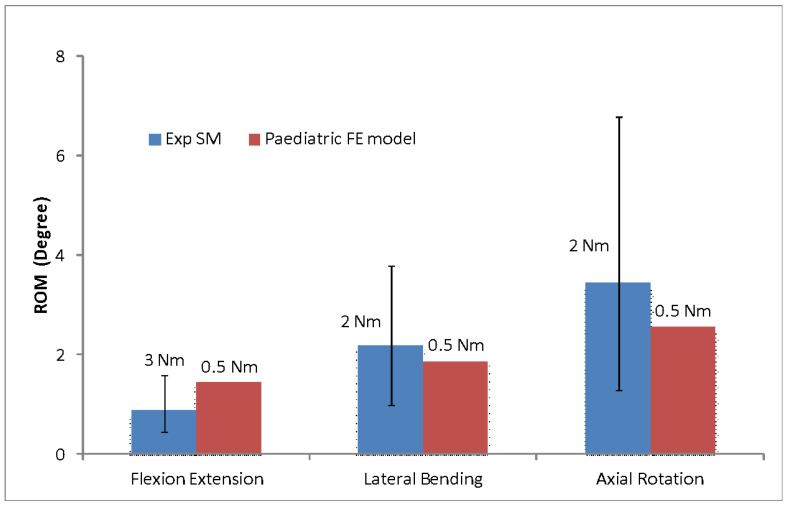
Studies on paediatric spines commonly use human adult or immature porcine spines as specimens, because it is difficult to obtain actual paediatric specimens. There are quite obvious differences, such as geometry, size, bone morphology, and orientation of facet joint for these specimens, compared to paediatric spine. Hence, development of synthetic models that can behave similarly to actual paediatric spines, particularly in term of range of motion (ROM), could provide a significant contribution for paediatric spine research. This study aims to develop a synthetic paediatric spine using finite element modelling and evaluate the reliability of the model by comparing it with the experimental data under certain load conditions. The ROM of the paediatric spine was measured using a validated FE model at ±0.5 Nm moment in order to determine the moment required by the synthetic spine to achieve the same ROM. The results showed that the synthetic spine required two moments, ±2 Nm for lateral-bending and axial rotation, and ±3 Nm for flexion-extension, to obtain the paediatric ROM. The synthetic spine was shown to be stiffer in flexion-extension but more flexible in lateral bending than the paediatric FE model, possibly as a result of the intervertebral disc's simplified shape and the disc's weak bonding with the vertebrae. Nevertheless, the synthetic paediatric spine has promising potential in the future as an alternative paediatric spine model for biomechanical investigation of paediatric cases.
Keywords: finite element analysis; paediatric FE model; range of motion; synthetic paediatric spine.
Conflict of interest statement
The authors declare no conflict of interest.
Figures


Similar articles
-
Superior-segment Bilateral Facet Violation in Lumbar Transpedicular Fixation, Part III: A Biomechanical Study of Severe Violation.Spine (Phila Pa 1976). 2020 May 1;45(9):E508-E514. doi: 10.1097/BRS.0000000000003327. Spine (Phila Pa 1976). 2020. PMID: 31770344
-
Incorporating ligament laxity in a finite element model for the upper cervical spine.Spine J. 2017 Nov;17(11):1755-1764. doi: 10.1016/j.spinee.2017.06.040. Epub 2017 Jun 30. Spine J. 2017. PMID: 28673824
-
The effect of follower load on the range of motion, facet joint force, and intradiscal pressure of the cervical spine: a finite element study.Med Biol Eng Comput. 2020 Aug;58(8):1695-1705. doi: 10.1007/s11517-020-02189-7. Epub 2020 May 28. Med Biol Eng Comput. 2020. PMID: 32462554
-
Biomechanical Effect of L4 -L5 Intervertebral Disc Degeneration on the Lower Lumbar Spine: A Finite Element Study.Orthop Surg. 2020 Jun;12(3):917-930. doi: 10.1111/os.12703. Epub 2020 May 31. Orthop Surg. 2020. PMID: 32476282 Free PMC article.
-
Experimental Analysis of Fabricated Synthetic Midthoracic Paediatric Spine as Compared to the Porcine Spine Based on Range of Motion (ROM).Appl Bionics Biomech. 2021 Sep 24;2021:2799415. doi: 10.1155/2021/2799415. eCollection 2021. Appl Bionics Biomech. 2021. PMID: 34608402 Free PMC article.
References
-
- Shiba K., Taneichi H., Namikawa T., Inami S., Takeuchi D., Nohara Y. Osseointegration improves bone–implant interface of pedicle screws in the growing spine: A biomechanical and histological study using an in vivo immature porcine model. Eur. Spine J. 2017;26:2754–2762. doi: 10.1007/s00586-017-5062-2. - DOI - PubMed
Grants and funding
LinkOut - more resources
Full Text Sources
