. 2023 Jun 22;24(13):10475.
doi: 10.3390/ijms241310475.

Improving Soluble Expression of SARS-CoV-2 Spike Priming Protease TMPRSS2 with an Artificial Fusing Protein

Affiliations

Improving Soluble Expression of SARS-CoV-2 Spike Priming Protease TMPRSS2 with an Artificial Fusing Protein

Xiao Ye et al. Int J Mol Sci. .

Abstract

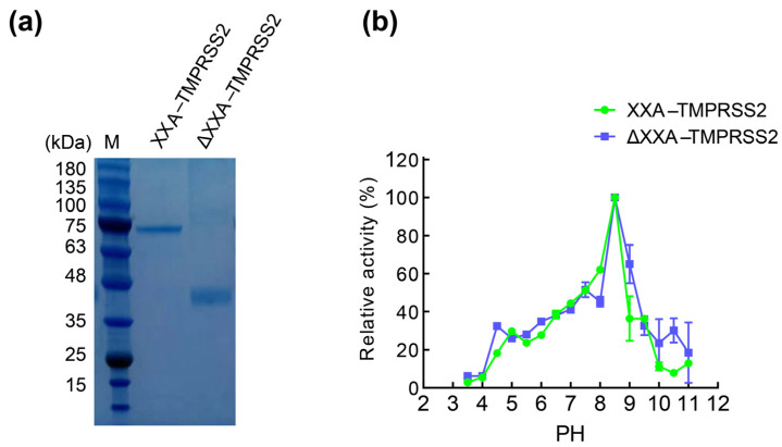
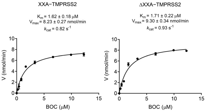
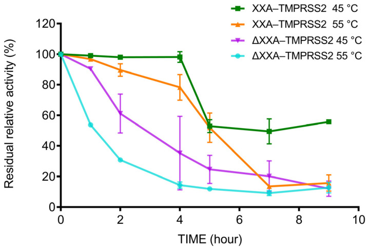
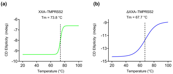
SARS-CoV-2 relies on the recognition of the spike protein by the host cell receptor ACE2 for cellular entry. In this process, transmembrane serine protease 2 (TMPRSS2) plays a pivotal role, as it acts as the principal priming agent catalyzing spike protein cleavage to initiate the fusion of the cell membrane with the virus. Thus, TMPRSS2 is an ideal pharmacological target for COVID-19 therapy development, and the effective production of high-quality TMPRSS2 protein is essential for basic and pharmacological research. Unfortunately, as a mammalian-originated protein, TMPRSS2 could not be solubly expressed in the prokaryotic system. In this study, we applied different protein engineering methods and found that an artificial protein XXA derived from an antifreeze protein can effectively promote the proper folding of TMPRSS2, leading to a significant improvement in the yield of its soluble form. Our study also showed that the fused XXA protein did not influence the enzymatic catalytic activity; instead, it greatly enhanced TMPRSS2's thermostability. Therefore, our strategy for increasing TMPRSS2 expression would be beneficial for the large-scale production of this stable enzyme, which would accelerate aniti-SARS-CoV-2 therapeutics development.

Keywords: SARS-CoV-2; TMPRSS2; artificial protein; protease; protein expression.


Conflict of interest statement

The authors declare no conflict of interest.

Figures

Figure 1
Figure 1
TMPRSS2 expressed as inclusion body. E. coli BL21 (DE3) was transfected with pET28a–His−TMPRSS2 and induced for 16 h with 0.6 or 1 mM IPTG at 16 or 25 °C. The cellular extract was separated into soluble supernatant and insoluble pellet fractions and analyzed via SDS–PAGE. The black arrow indicates the recombinant protein His−TMPRSS2.
Figure 2
Figure 2
XXA tag promotes soluble expression of TMPRSS2. E. coli BL21(DE3) transfected with pET–His–TM, pET–MBP–TM, pET–SUMO–TM, pGEX–GST–TM, (a) and pET–XXA–TMPRSS2; (b) the above TM refers to TMPRSS2 and then induced with IPTG (1 mM) for 16 h at 16 °C. The soluble cell lysate (S) and insoluble pellets (P) were separated via centrifugation at 8000 rpm. pET–XXA–TMPRSS2 transfected cells without IPTG induction were chosen as negative control (C). The red box highlights the cell lysate from cells transfected with each plasmid, and the black arrows indicate the recombinant TMPRSS2 protein. (c) The proteolytic activity was examined with the crude soluble cell lysates using fluorescent trypsin substrate [Boc–Gln–Ala–Arg–AMC]. The relative activity of the enzyme was calculated considering the maximum activity as 100%. The error bar represents the standard deviation of two independent assays.
Figure 3
Figure 3
Purification of XXA tagged TMRPPSS2. (a) SDS–PAGE analysis of different samples taken during the purification process of the recombinant protein. Lane M: protein standard molecular weight. Lane 1: crude soluble cell lysate containing XXA–TMPRSS2. Lane 2: flow–through fraction from the Ni–NTA affinity Sepharose column. Lanes 3 to 5: protein sample eluted by low concentration of Imidazole (20 μM). Lanes 6 to 9: purified protein eluted by high concentration of Imidazole (100–300 μM) from one Ni–NTA affinity Sepharose column. (b) Lanes 1 to 4 purified XXA–TMPRSS2 proteins (corresponding to lanes 6 to 9 in panel (a) were analyzed by immunoblotting with anti–His antibody.
Figure 4
Figure 4
The MS–MS fragmentation spectra of four peptides selected from the peptide mass fingerprint (PMF) spectrum. The result analysis was performed from fragments of XXA–TMPRSS2 derived via trypsin digestion. Representative sequences coverage of these fragments was highlighted in red. The amino acid sequence corresponding to protein XXA was indicated in black, while the sequence of TMPRSS2 was shown in purple. An HRV 3C protease–specific recognition sequence LEVLFQ/GP was introduced into the recombinant protein between XXA and TMPRSS2.
Figure 5
Figure 5
Effect of pH on the activity of XXA–TMPRSS2 and ΔXXA–TMPRSS2. (a): ΔXXA–TMPRSS2 was generated by the proteolytic cleavage of XXA–TMPRSS2 with HRV 3C Protease. (b): The influence of pH on the proteolytic activity of TMPRSS2 was shown. The reaction was carried out at different pH values ranging from 3.0 to 11.0 at room temperature with Boc–Gln–Ala–Arg–AMC as substrate. Values represent the mean ± standard deviation of three measurements. The relative activity of the enzyme was calculated considering the maximum activity as 100%.
Figure 6
Figure 6
Kinetic parameter for XXA–TMPRSS2 and ΔXXA–TMPRSS2. The Vmax, kcat, and Km were measured via plotting reaction velocity versus substrate Boc–Gln–Ala–Arg–AMC concentration at room temperature in pH 8.0. Values represent the mean ± standard deviation of three measurements.
Figure 7
Figure 7
Thermostability assay of XXA–TMPRSS2 and ΔXXA–TMPRSS2. Thermostability of recombinant XXA–TMPRSS2 and ΔXXA–TMPRSS2 was determined by preincubating 1 mg/mL at 45 and 55 °C for designated time periods and then assaying the activity in reaction buffer at room temperature as described in “Materials and Methods”. The relative activity of the enzyme was calculated considering the maximum activity as 100%.
Figure 8
Figure 8
Protein denaturation analysis of XXA−TMPRSS2 and ΔXXA−TMPRSS2. (a) CD measurements at 220 nm of XXA−TMPRSS2 at different temperatures (20−100 °C); (b) CD measurements at 220 nm of ΔXXA−TMPRSS2 at different temperatures (20−100 °C). Dashed lines indicate the melting temperatures (Tm or the transition midpoint) of each enzyme. The Tm represents the temperature at which 50% of the protein is unfolded, which corresponds to the midpoint of the sigmoidal unfolding curve.

Similar articles

References

    1. de Loyola M.B., Dos Reis T.T.A., de Oliveira G., da Fonseca Palmeira J., Arganaraz G.A., Arganaraz E.R. Alpha–1–antitrypsin: A possible host protective factor against COVID-19. Rev. Med. Virol. 2021;31:e2157. doi: 10.1002/rmv.2157. - DOI - PMC - PubMed
    1. Hoffmann M., Kleine–Weber H., Schroeder S., Kruger N., Herrler T., Erichsen S., Schiergens T.S., Herrler G., Wu N.H., Nitsche A., et al. SARS-CoV-2 Cell Entry Depends on ACE2 and TMPRSS2 and Is Blocked by a Clinically Proven Protease Inhibitor. Cell. 2020;181:271–280.e8. doi: 10.1016/j.cell.2020.02.052. - DOI - PMC - PubMed
    1. Wang M.Y., Zhao R., Gao L.J., Gao X.F., Wang D.P., Cao J.M. SARS-CoV-2: Structure, Biology, and Structure–Based Therapeutics Development. Front. Cell. Infect. Microbiol. 2020;10:587269. doi: 10.3389/fcimb.2020.587269. - DOI - PMC - PubMed
    1. Walls A.C., Park Y.J., Tortorici M.A., Wall A., McGuire A.T., Veesler D. Structure, Function, and Antigenicity of the SARS-CoV-2 Spike Glycoprotein. Cell. 2020;183:1735. doi: 10.1016/j.cell.2020.11.032. - DOI - PMC - PubMed
    1. Papa G., Mallery D.L., Albecka A., Welch L.G., Cattin–Ortola J., Luptak J., Paul D., McMahon H.T., Goodfellow I.G., Carter A., et al. Furin cleavage of SARS-CoV-2 Spike promotes but is not essential for infection and cell–cell fusion. PLoS Pathog. 2021;17:e1009246. doi: 10.1371/journal.ppat.1009246. - DOI - PMC - PubMed

Substances