. 2023 Jun 23;24(13):10527.
doi: 10.3390/ijms241310527.

Microcystin-LR-Induced Interaction between M2 Tumor-Associated Macrophage and Colorectal Cancer Cell Promotes Colorectal Cancer Cell Migration through Regulating the Expression of TGF-β1 and CST3

Affiliations

Microcystin-LR-Induced Interaction between M2 Tumor-Associated Macrophage and Colorectal Cancer Cell Promotes Colorectal Cancer Cell Migration through Regulating the Expression of TGF-β1 and CST3

Xinying Jiang et al. Int J Mol Sci. .

Abstract

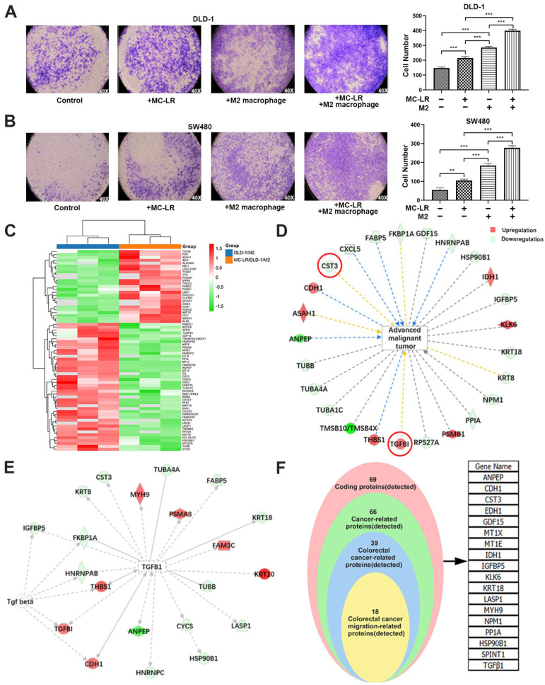
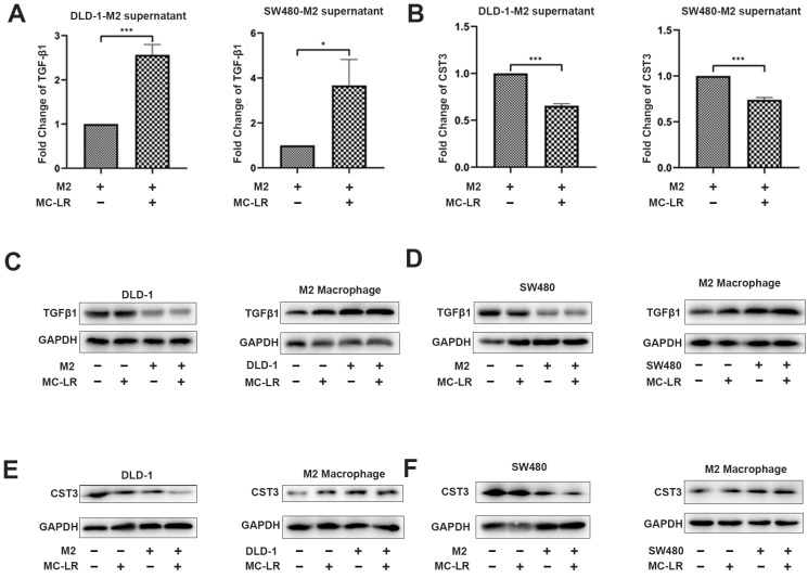
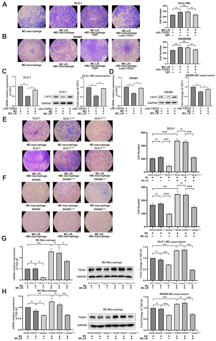
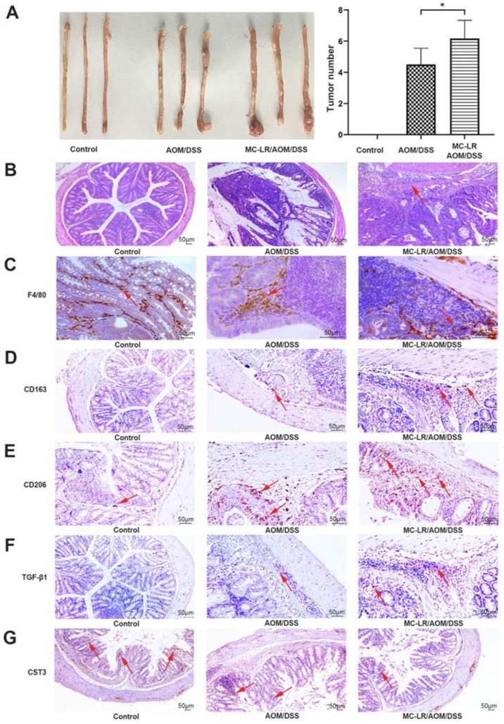
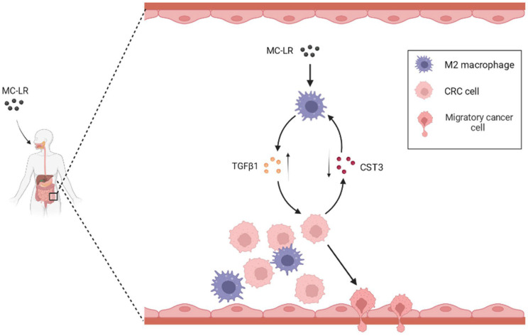
Microcystin-LR (MC-LR) is a toxic secondary metabolite produced by cyanobacteria that has been demonstrated to promote colorectal cancer (CRC). However, the mechanism by which MC-LR enhances CRC in the tumor microenvironment (TME) is poorly understood. To elucidate its role in TME, a co-culture system was established using CRC cells and M2 macrophages in a Transwell chamber. The study found that MC-LR promotes CRC cell migration by upregulating TGF-β1 expression and secretion in M2 macrophages and downregulating CST3 in CRC cells. Neutralizing TGF-β1 increased CST3 expression in CRC cells, while overexpressing CST3 in CRC cells suppressed TGF-β1 expression in M2 macrophages, both of which weakened MC-LR-induced cellular motility in the co-culture system. In vivo, the mice in the MC-LR/AOM/DSS group had more tumor nodules, deeper tumor invasion, and higher M2 macrophage infiltration compared to the AOM/DSS group, and the expression of TGF-β1 and CST3 in tumors was consistent with the cellular level. Overall, this study provides insights into the regulatory mechanism of MC-LR on TME, revealing that MC-LR upregulates the expression and secretion of TGF-β1 in M2 macrophages, which in turn inhibits the expression of CST3 in CRC cells to promote migration.

Keywords: M2 tumor-associated macrophages; MC-LR; TGF-β1; colorectal cancer; migration.


Conflict of interest statement

The authors declare no conflict of interest.

Figures

Figure 1
Figure 1
MC-LR-induced CRC cell–M2 macrophage interaction promoted CRC cell migration and analysis of relative proteins. The effect of MC-LR on the migration of (A) DLD-1 and (B) SW480 cells was determined by Transwell. The migration cells were stained with crystal violet, and the pictures were captured under light microscopy (×40). Secreted proteins were detected by iTRAQ quantitative proteomics technology. Samples were harvested from DLD-1 cell–M2 macrophage co-culture system with or without 25 nM MC-LR treatment for 48 h. (C) Differential expression proteins in co-culture system were colored based on the heat map scale (upregulated: red, FC ≥ 1.2; downregulated: blue, FC ≤ −1.2) by iTRAQ quantitative proteomics technology. IPA was used to analyze (D) the relationship between advanced malignant tumors and screened proteins, and (E) the interaction between TGF-β1 and related proteins. Red represents increased proteins; and green represents decreased proteins. (F) The proteins related to the migration of CRC cells were screened out. All data were presented as the mean ± SD from at least three independent experiments. ** p < 0.01, and *** p < 0.001, compared with the control group.
Figure 2
Figure 2
MC-LR promotes the expression of TGF-β1 in M2 macrophages and inhibits the expression of CST3 in CRC cells under the co-culture system. (A) Secretion of TGF-β1 was detected by ELISA in the supernatant of co-culture system with 25 nM MC-LR for 48 h. (B) Secretion of CST3 was detected by ELISA in the supernatant of co-culture system with 25 nM MC-LR for 48 h. Protein expression of TGF-β1 and CST3 in (C,E) DLD-1 cell–M2 macrophage and (D,F) SW480 cell–M2 macrophage co-culture system was detected by Western blotting, respectively. All data were presented as the mean ± SD from at least three independent experiments. * p < 0.05, and *** p < 0.001, compared with the control group.
Figure 3
Figure 3
MC-LR prompted CRC cell migration by regulating the expression of TGF-β1 and CST3 in the co-culture system. The migration of (A,B) CRC cells in the co-culture system. Migration of (A) DLD-1 cells and (B) SW480 cells in the co-culture system which were treated with 0.8 μg/mL TGF-β1-neutralizing antibody and 25 nM MC-LR. IgG was used as a negative control. The pictures were captured under light microscopy (×40). mRNA and protein expression and secretion of CST3 in (C) DLD-1 cell and (D) SW480 cells -M2 macrophage co-culture systems which were treated with 25 nM MC-LR or 25 nM MC-LR/TGF-β1-neutralizing antibody (0.8 μg/mL) for 48 h, detected by qPCR, Western blotting, and ELISA, respectively. The migration of (E) DLD-1 and (F) SW480 cells with overexpressing CST3 which were co-cultured with M2 macrophages and MC-LR is at the concentration of 25 nM for 48 h. The pictures were captured under light microscopy (×40). mRNA and protein expression and secretion of TGF-β1 in M2 macrophages in (G) DLD-1 cells and (H) SW480 cell–M2 macrophage, detected by qPCR, Western blotting, and ELISA. All mRNA expression levels were calculated by the double delta CT method and normalized to the expression of GAPDH. All data were presented as the mean ± SD from at least three independent experiments. * p < 0.05, ** p < 0.01, and *** p < 0.001, ns present no significant, compared with the control group.
Figure 4
Figure 4
MC-LR increased M2 macrophage infiltration and decreased the expression of CST3 to exacerbate tumor progression in AOM/DSS-model mice. (A) Gross macroscopic images of the colorectal tissues of mice in each group were represented (n = 10). The number of colorectal tumors in mice was counted. (B) Colorectal tumor tissues of mice were stained by H&E and the arrow indicates tumor infiltration (scale bar: 50 μm). Data were presented as the means ± SD (n = 10). * p < 0.05, compared with the control group. The IHC results of CRC tissues were shown in (CF), and (G), which represent the expression of F4/80, CD163, CD206, TGF-β1, and CST3, respectively, and the arrows indicated the positive expression of corresponding measurement indicators (scale bar: 50 μm). Sequential representation: the control and the AOM/DSS group were treated with saline; and MC-LR/AOM/DSS group was treated with 40 μg/kg body weight MC-LR daily for 15 days.
Figure 5
Figure 5
The mechanism of the promoted migration of CRC cells induced by MC-LR in the TME.

Similar articles

Cited by

References

    1. Ding Q., Liu K., Song Z., Sun R., Zhang J., Yin L., Pu Y. Effects of Microcystin-LR on Metabolic Functions and Structure Succession of Sediment Bacterial Community under Anaerobic Conditions. Toxins. 2020;12:183. - PMC - PubMed
    1. MacKintosh C., Beattie K.A., Klumpp S., Cohen P., Codd G.A. Cyanobacterial microcystin-LR is a potent and specific inhibitor of protein phosphatases 1 and 2A from both mammals and higher plants. FEBS Lett. 1990;264:187–192. - PubMed
    1. Laureano-Rosario A.E., McFarland M., Bradshaw D.J., Metz J., Brewton R.A., Pitts T., Perricone C., Schreiber S., Stockley N., Wang G., et al. Dynamics of microcystins and saxitoxin in the Indian River Lagoon, Florida. Harmful Algae. 2021;103:102012. - PubMed
    1. Xu Q., Chen W., Gao G. Seasonal variations in microcystin concentrations in Lake Taihu, China. Environ. Monit. Assess. 2008;145:75–79. - PubMed
    1. Lone Y., Koiri R.K., Bhide M. An overview of the toxic effect of potential human carcinogen Microcystin-LR on testis. Toxicol. Rep. 2015;2:289–296. doi: 10.1016/j.toxrep.2015.01.008. - DOI - PMC - PubMed

Substances