. 2023 Jun 24;24(13):10577.
doi: 10.3390/ijms241310577.

Molecular Dynamics Simulations of Matrix Metalloproteinase 13 and the Analysis of the Specificity Loop and the S1'-Site

Affiliations

Molecular Dynamics Simulations of Matrix Metalloproteinase 13 and the Analysis of the Specificity Loop and the S1'-Site

Jun Yong Choi et al. Int J Mol Sci. .

Abstract

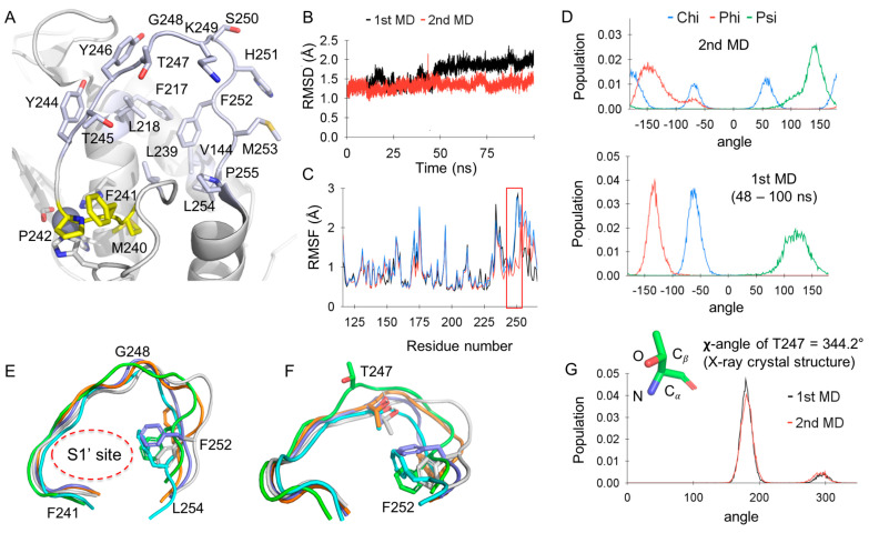
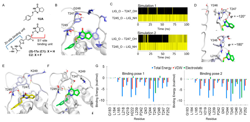
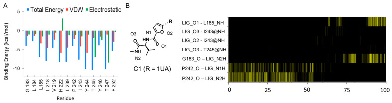
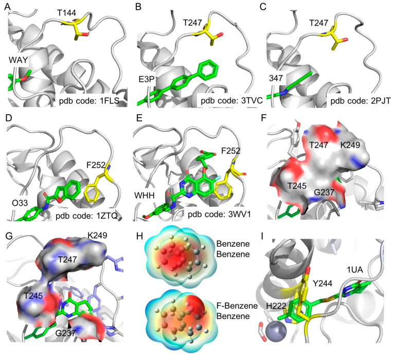
The specificity loop of Matrix Metalloproteinases (MMPs) is known to regulate recognition of their substrates, and the S1'-site surrounded by the loop is a unique place to address the selectivity of ligands toward each MMP. Molecular dynamics (MD) simulations of apo-MMP-13 and its complex forms with various ligands were conducted to identify the role of the specificity loop for the ligand binding to MMP-13. The MD simulations showed the dual role of T247 as a hydrogen bond donor to the ligand, as well as a contributor to the formation of the van der Waal surface area, with T245 and K249 on the S1'-site. The hydrophobic surface area mediated by T247 blocks the access of water molecules to the S1'-site of MMP-13 and stabilizes the ligand in the site. The F252 residue is flexible in order to search for the optimum location in the S1'-site of the apo-MMP-13, but once a ligand binds to the S1'-site, it can form offset π-π or edge-to-π stacking interactions with the ligand. Lastly, H222 and Y244 provide the offset π-π and π-CH(Cβ) interactions on each side of the phenyl ring of the ligand, and this sandwiched interaction could be critical for the ligand binding to MMP-13.

Keywords: MD simulations; S1′−site; matrix metalloproteinase 13; selective MMP inhibitors; specificity loop; π−CH(Cβ) interactions.


Conflict of interest statement

The authors declare no conflict of interest. The funders had no role in the design of the study; in the collection, analyses, or interpretation of data; in the writing of the manuscript; or in the decision to publish the results.

Figures

Figure 1
Figure 1
MD simulations of the apo−MMP−13 structure: (A) The specificity loop and S1′−site of MMP-13; Residues in the specificity loop and S1′−site are light blue, and residues in the Met−turn are yellow; (B) RMSD of MD simulations of apo−MMP-13; (C) RMSF of all residues in MD run 1 (blue), run 1 (48–100 ns, red), and run 2 (black); (D) Dihedral angle analysis of F252: (top) MD run 2 (0–100 ns) and (bottom) MD run 1 (48–100 ns); (E) The specificity loops of MMP−13 from MD simulations; the green loop corresponds to the X-ray crystal structure (PDB code: 4L19).; (F) The conformation of T247 and F252 from the MD simulations of MMP-13; the green loop corresponds to the X-ray crystal structure (PDB code: 4L19).; (G) The χ−angle histogram of T247 from the MD simulations (20–100 ns) and the χ−angle of T247 in the X-ray co−crystal structure.
Figure 2
Figure 2
MD simulations of the MMP−13 − 1UA, C1, or C2 complex: (A) The structures of 1UA, C1, and C2; (B) The open conformation of the specificity loop of MMP−13; (C) The H−bond histogram analysis of MMP−13 − 1UA complex; simulation 1 (binding pose 1) and simulation 2 (binding pose 2). The yellow line represents the presence of the H−bond interaction at each time point.; LIG_O and LIG_NH stand for carbonyl oxygen and NH of 1UA, and T245_O and T247_OH for oxygen of the T245 amide backbone and OH of the T247 residue, respectively; (D) Two different conformations of T247 depending on the φ angle of Y246; (E) The binding pose 1 of 1UA; (F) The binding pose 2 of 1UA; (G) The per−residue energy decomposition analyses of MMP−13 − 1UA complex; the binding pose 1 (left) and the binding pose 2 (right).
Figure 3
Figure 3
Analysis of MD simulations of MMP−13 − C1 complex: (A) The per−residue energy decomposition analyses of MMP−13 − C1 complex; (B) The H−bond histogram analysis of MMP−13 − C1 complex; the yellow line represents the presence of the H−bond interaction at each time point. Only the H−bond interactions in the Zn−binding site are present.; LIG_O1, O2, and O3 stand for oxygen of C1, and LIG_N1H and N2H for hydrogen on the amide unit of C1 (left panel). L185_NH/I243_NH/T245_NH and G183_O/P242_O stand for hydrogen and oxygen on the amide backbone of each amino acid, respectively.
Figure 4
Figure 4
Analysis of MD simulations of MMP−13 complexed with various ligands occupying S1′-site: (A) Structures of ligands used for MD simulations; af are scaffolds of ligands, and the complete 2−D structures of ligands are present in Table S4.; (B) A data plot of pIC50 vs. binding affinities calculated from MM/GBSA; (C) RMSD of MD simulations of MMP−13 complexed with C99, C100, C101, and C102; (D) The offset π−π stacking interactions of benzene and fluorobenzene obtained from QM calculations; (E) The F252 energy decomposition analyses of MMP−13 complexed with C99−C110 via MM/GBSA calculations; (F) The shift of C103 (green) in the S1′−site due to additional H−bond interactions.
Figure 5
Figure 5
The specificity loop and S1′−site of MMP−13 complexed with ligands: (AC) The NMR structure of MMP−13 (pdb code: 1FM1) and the X-ray co-crystal structures of MMP−13 (pdb codes: 3TVC and 2PJT), respectively, which shows the flexible conformation of T144/T247 (yellow). The part of each ligand in the S1′-site is shown in green, and each ligand code is labeled; (D,E) The edge−to−face and offset π−π stacking interactions between F252 and ligands in the S1′−site of MMP−13, respectively; (F) The S1′−site closed by the hydrophobic surface made by G237, T245, T247, and K249 in the specificity loop; (G) The open state of S1′−site; (H) The electrostatic potential surface of the offset π–π stacking interaction of benzene and fluorobenzene; (I) The sandwiched π−π and π−CH (Cβ) interactions by the ligand, H222, and Y244, which could be critical for the ligand binding to MMP−13.

Similar articles

Cited by

References

    1. de Almeida L.G.N., Thode H., Eslambolchi Y., Chopra S., Young D., Gill S., Devel L., Dufour A. Matrix Metalloproteinases: From Molecular Mechanisms to Physiology, Pathophysiology, and Pharmacology. Pharmacol. Rev. 2022;74:712–768. doi: 10.1124/pharmrev.121.000349. - DOI - PubMed
    1. Winkler J., Abisoye-Ogunniyan A., Metcalf K.J., Werb Z. Concepts of extracellular matrix remodelling in tumour progression and metastasis. Nat. Commun. 2020;11:5120. doi: 10.1038/s41467-020-18794-x. - DOI - PMC - PubMed
    1. Diaz-Canestro C., Puspitasari Y.M., Liberale L., Guzik T.J., Flammer A.J., Bonetti N.R., Wust P., Costantino S., Paneni F., Akhmedov A., et al. MMP-2 knockdown blunts age-dependent carotid stiffness by decreasing elastin degradation and augmenting eNOS activation. Cardiovasc. Res. 2022;118:2385–2396. doi: 10.1093/cvr/cvab300. - DOI - PubMed
    1. Visse R., Nagase H. Matrix metalloproteinases and tissue inhibitors of metalloproteinases: Structure, function, and biochemistry. Circ. Res. 2003;92:827–839. doi: 10.1161/01.RES.0000070112.80711.3D. - DOI - PubMed
    1. Fields G.B. The Rebirth of Matrix Metalloproteinase Inhibitors: Moving Beyond the Dogma. Cells. 2019;8:984. doi: 10.3390/cells8090984. - DOI - PMC - PubMed

MeSH terms

Substances