. 2023 Jun 25;24(13):10632.
doi: 10.3390/ijms241310632.

ECM Composition Differentially Regulates Intracellular and Extracellular pH in Normal and Cancer Pancreatic Duct Epithelial Cells

Affiliations

ECM Composition Differentially Regulates Intracellular and Extracellular pH in Normal and Cancer Pancreatic Duct Epithelial Cells

Daria Di Molfetta et al. Int J Mol Sci. .

Abstract

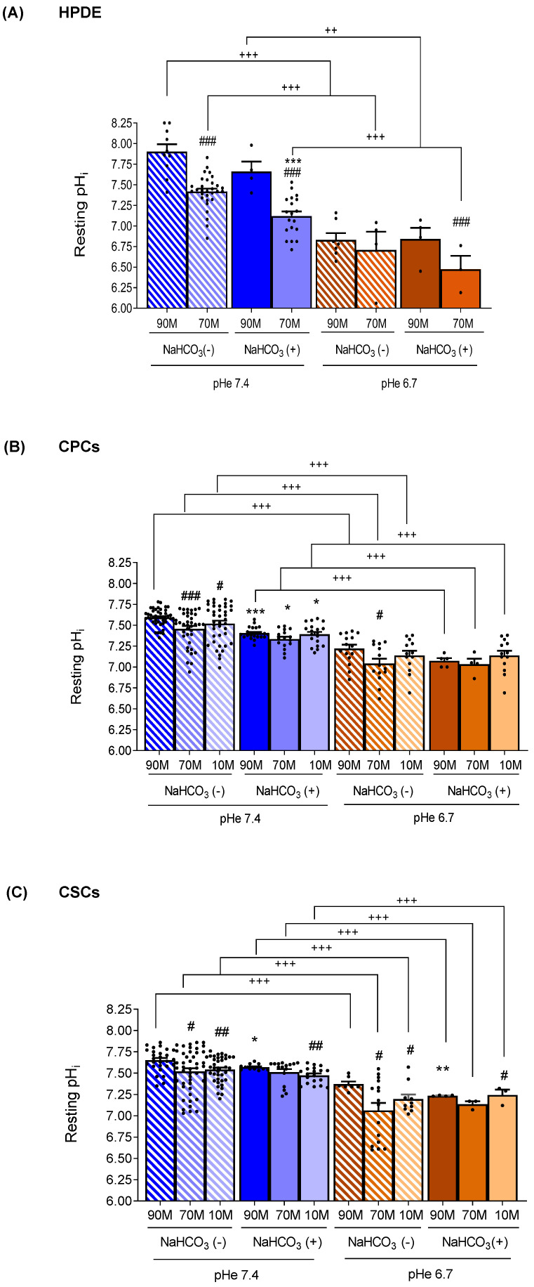
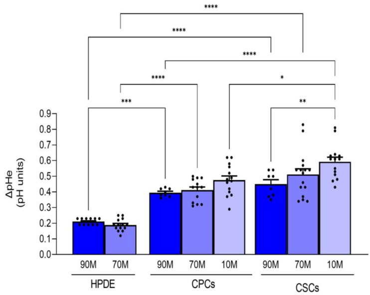
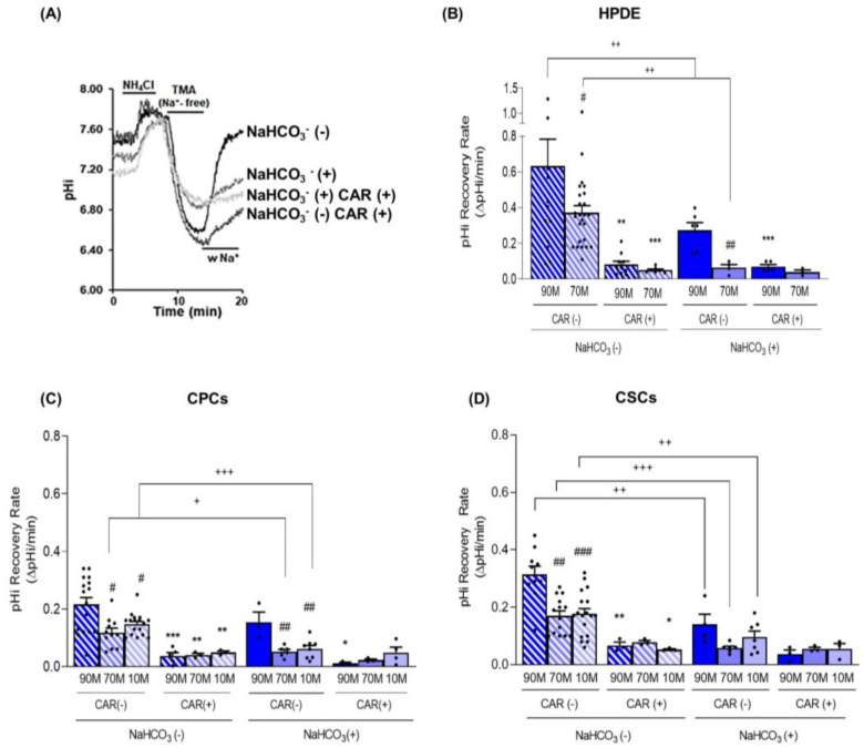
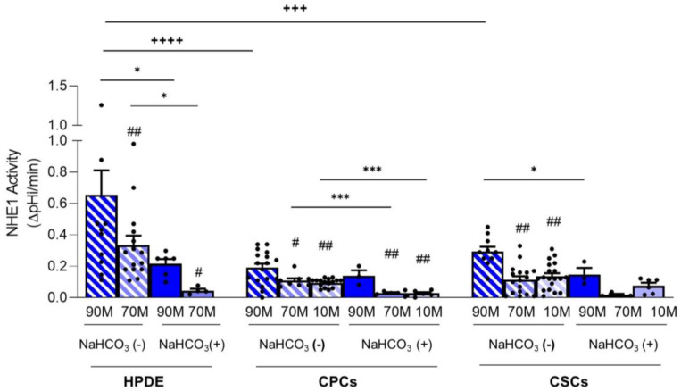
Intracellular pH (pHi) regulation is a challenge for the exocrine pancreas, where the luminal secretion of bicarbonate-rich fluid is accompanied by interstitial flows of acid. This acid-base transport requires a plethora of ion transporters, including bicarbonate transporters and the Na+/H+ exchanger isoform 1 (NHE1), which are dysregulated in Pancreatic Ductal Adenocarcinoma (PDAC). PDAC progression is favored by a Collagen-I rich extracellular matrix (ECM) which exacerbates the physiological interstitial acidosis. In organotypic cultures of normal human pancreatic cells (HPDE), parenchymal cancer cells (CPCs) and cancer stem cells (CSCs) growing on matrices reproducing ECM changes during progression, we studied resting pHi, the pHi response to fluxes of NaHCO3 and acidosis and the role of NHE1 in pHi regulation. Our findings show that: (i) on the physiological ECM, HPDE cells have the most alkaline pHi, followed by CSCs and CPCs, while a Collagen I-rich ECM reverses the acid-base balance in cancer cells compared to normal cells; (ii) both resting pHi and pHi recovery from an acid load are reduced by extracellular NaHCO3, especially in HPDE cells on a normal ECM; (iii) cancer cell NHE1 activity is less affected by NaHCO3. We conclude that ECM composition and the fluctuations of pHe cooperate to predispose pHi homeostasis towards the presence of NaHCO3 gradients similar to that expected in the tumor.

Keywords: NHE1; PDAC; bicarbonate transport.


Conflict of interest statement

The authors declare no conflict of interest. The funders had no role in the design of the study; in the collection, analyses, or interpretation of data; in the writing of the manuscript, or in the decision to publish the results.

Figures

Figure 1
Figure 1
Effect of the ECM composition (from a less fibrotic to a more fibrotic ECM) and different pHe (pHe 7.4 and pHe 6.7) in the presence (NaHCO3 (+)) or absence of NaHCO3 (NaHCO3 (−)) on resting pHi. The cells were alternatively perfused with Ringer with and without NaHCO3 (see Supplementary Table S1) at both pHes to determine the effect of the changing pHe and the presence or absence of NaHCO3 on resting pHi. (a) The normal HPDE cells (A) were grown on a matrix resembling the normal pancreatic ECM (90M-10C) and a low fibrotic ECM (70M-30C), while the tumor cells (B) CPCs and (C) CSCs, concordant with the increasing level of desmoplasia in fully established PDAC, were also grown on a highly fibrotic ECM (10M-90C). Significance between groups: # p < 0.05; ## p < 0.01; ### p < 0.001 compared to 90M in the same NaHCO3/pHe conditions; * p < 0.05; ** p < 0.01; *** p < 0.001 comparing NaHCO3 (+) versus NaHCO3 (−) on the same ECM/pHe; ++ p < 0.01; +++ p < 0.001.
Figure 2
Figure 2
Effect of the ECM composition (from a less fibrotic to a more fibrotic ECM) on the ability of the three cell lines to acidify their extracellular NaHCO3-containing medium measured with single-barreled H+-sensitive microelectrodes. Acidification is expressed as ∆pH in the medium of confluent cells over 24 h. The acidification rate of the HPDE cells was significantly less than either the CPCs or the CSCs on all ECMs. Significance between groups: * p < 0.05; ** p < 0.01; *** p < 0.001 and **** p < 0.0001.
Figure 3
Figure 3
(A) Representative traces of the pHi recovery rate from an NH4Cl prepulse-induced acidification in HPDE 3D organotypic cultures labelled with the pH-sensitive probe BCECF. The experiment was performed according to the Boron protocol (please see Materials and Methods) with or without NaHCO3 ((NaHCO3 (+) or NaHCO3 (−), respectively), and in the absence (CAR (−)) and presence (CAR (+)) of Cariporide (5 µM) during the pH recovery to evaluate the contribution of NHE1. In both the normal HPDE cells (B) and in the cancer cells, CPCs (C) and CSCs (D), the pHi recovery is always dependent on NHE1 activity in the absence of NaHCO3. Results are presented as mean ± SEM. Significance between groups: * p < 0.05; ** p < 0.01; *** p < 0.001 compared to their own pHi measured without Cariporide on the same ECM; # p < 0.05; ## p < 0.01; ### p < 0.001 compared to 90M-10C for each cell line; + p < 0.05; ++ p < 0.01; +++ p < 0.001.
Figure 4
Figure 4
NHE1 activity calculated as the pHi recovery in control condition subtracted from pHi recovery in the presence of Cariporide for HPDE, CPC and CSC cell lines. Significance between groups: # p < 0.05; ## p < 0.01 compared to 90M-10C for each cell line; * p < 0.05, *** p < 0.001; +++ p < 0.001 and ++++ p < 0.0001.

Similar articles

Cited by

References

    1. Stark A., Eibl G. Pancreatic Ductal Adenocarcinoma. Pancreapedia: The Exocrine Pancreas Knowledge Base. 2015. [(accessed on 23 May 2015)]. Available online: https://pancreapedia.org/?q=node/9002.
    1. Hezel A.F., Kimmelman A.C., Stanger B.Z., Bardeesy N., DePinho R.A. Genetics and Biology of Pancreatic Ductal Adenocarcinoma. Genes. Dev. 2006;20:1218–1249. doi: 10.1101/gad.1415606. - DOI - PubMed
    1. Lee M.G., Ohana E., Park H.W., Yang D., Muallem S. Molecular Mechanism of Pancre-Atic and Salivary Gland Fluid and HCO 3 Secretion. Physiol. Rev. 2012;92:39–74. doi: 10.1152/physrev.00011.2011. - DOI - PMC - PubMed
    1. Dahl-Jensen S.B., Yennek S., Flasse L., Larsen H.L., Sever D., Karremore G., Novak I., Sneppen K., Grapin-Botton A. Deconstructing the Principles of Ductal Network Formation in the Pancreas. PLoS Biol. 2018;16:e2002842. doi: 10.1371/journal.pbio.2002842. - DOI - PMC - PubMed
    1. Hegyi P., Maléth J., Venglovecz V., Rakonczay Z. Pancreatic Ductal Bicarbonate Secretion: Challenge of the Acinar Acid Load. Front. Physiol. 2011;2:36. doi: 10.3389/fphys.2011.00036. - DOI - PMC - PubMed