. 2023 Jun 25;24(13):10627.
doi: 10.3390/ijms241310627.

Cross-Linking Mass Spectrometry on P-Glycoprotein

Affiliations

Cross-Linking Mass Spectrometry on P-Glycoprotein

Gabriella Gellen et al. Int J Mol Sci. .

Abstract

The ABC transporter P-glycoprotein (Pgp) has been found to be involved in multidrug resistance in tumor cells. Lipids and cholesterol have a pivotal role in Pgp's conformations; however, it is often difficult to investigate it with conventional structural biology techniques. Here, we applied robust approaches coupled with cross-linking mass spectrometry (XL-MS), where the natural lipid environment remains quasi-intact. Two experimental approaches were carried out using different cross-linkers (i) on living cells, followed by membrane preparation and immunoprecipitation enrichment of Pgp, and (ii) on-bead, subsequent to membrane preparation and immunoprecipitation. Pgp-containing complexes were enriched employing extracellular monoclonal anti-Pgp antibodies on magnetic beads, followed by on-bead enzymatic digestion. The LC-MS/MS results revealed mono-links on Pgp's solvent-accessible residues, while intraprotein cross-links confirmed a complex interplay between extracellular, transmembrane, and intracellular segments of the protein, of which several have been reported to be connected to cholesterol. Harnessing the MS results and those of molecular docking, we suggest an epitope for the 15D3 cholesterol-dependent mouse monoclonal antibody. Additionally, enriched neighbors of Pgp prove the strong connection of Pgp to the cytoskeleton and other cholesterol-regulated proteins. These findings suggest that XL-MS may be utilized for protein structure and network analyses in such convoluted systems as membrane proteins.

Keywords: P-glycoprotein; cancer; cholesterol; cross-linking mass spectrometry; membrane; mono-link; multidrug resistance; protein structure.


Conflict of interest statement

The authors declare no conflict of interest. The funders had no role in the design of the study; in the collection, analyses, or interpretation of data; in the writing of the manuscript; or in the decision to publish the results.

Figures

Figure 1
Figure 1
Regions of Pgp on its primary and tertiary structures. (A) Primary structure of Pgp visualized using Protter 1.0 [22]. Regions of the protein indicated in pink form nucleotide-binding site 1 (NBS1) and the ones indicated in brown belong to NBS2. TMH 1–12 are indicated in blue. Cholesterols identified in the 6qex PDB structure are labeled in yellow. (B) Tertiary structure of Pgp using inward-facing human Pgp structure, 6qex, with its specific regions indicated. TMD1 (light blue) connected to NBD1 (dark blue), TMD2 (pale green) connected to NBD2 (dark green), NBS1 regions (pink), NBS2 (brown) and flexible linker and elbow helices (grey). N-glycans on ECL1 are blue, while oxygen atoms are labeled in red (see abbreviations in the text of the Introduction).
Figure 1
Figure 1
Regions of Pgp on its primary and tertiary structures. (A) Primary structure of Pgp visualized using Protter 1.0 [22]. Regions of the protein indicated in pink form nucleotide-binding site 1 (NBS1) and the ones indicated in brown belong to NBS2. TMH 1–12 are indicated in blue. Cholesterols identified in the 6qex PDB structure are labeled in yellow. (B) Tertiary structure of Pgp using inward-facing human Pgp structure, 6qex, with its specific regions indicated. TMD1 (light blue) connected to NBD1 (dark blue), TMD2 (pale green) connected to NBD2 (dark green), NBS1 regions (pink), NBS2 (brown) and flexible linker and elbow helices (grey). N-glycans on ECL1 are blue, while oxygen atoms are labeled in red (see abbreviations in the text of the Introduction).
Figure 5
Figure 5
Solvent accessibility of lysines on Pgp. (A) Electrostatic potential surface representation of inward-facing Pgp (6qex); nitrogen atoms of all lysines are represented as grey balls, and buried ones are colored in yellow. Five lysines with a legend are the ones which were modified by cross-linkers. (B) Hydrophobicity and charge distribution on Pgp’s surface. Carbon atoms not bound to nitrogen or oxygen atoms are yellow, oxygens with negative charges in glutamate and aspartate are red and nitrogens carrying positive charges in lysine and arginine are blue, while all other atoms are white [51]. The inlet shows extracellular lysines of which only K734 and K967 are surface exposed; however, when Pgp is in complex with UIC2 or 15D3 they are also buried.
Figure 6
Figure 6
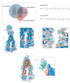
Mono-links and cross-links on Pgp. (A) Overlap of cross-links between different sample preparation approaches. Eleven cross-links were identified with all sample preparation approaches, with a 27% overlap between the living cell and the on-bead DSSO methods. No cross-links were identified with the living cell BS2Gd0/d4 approach. (B) Linear plots of cross-linked sites on human Pgp identified with on-bead and living cell approaches. All lysines are indicated in light blue, and cross-links are purple. (C) Mono-links highlighted in green balls and cross-links indicated by red lines on the structure of Pgp in complex with UIC2 antibody (6qex) based on the results of the cross-linking approaches. Lysines which are not mono-linked are depicted as grey balls, and cholesterols and lipids are yellow. The inlet shows the ICH3 loop involved in cross-links highlighted in pink, which was previously described to be influenced by the presence of cholesterol. (D) Mono-links of Walker A regions and other segments of nucleotide binding sites (NBS) emphasized in pink. (E) ICH3 directly connected to ECL5, cross-linked to C-loop, which has cross-link with ICH4, directly connected to ECL6. Connections of these regions on Pgp suggest a complex interplay between segments that have previously been described as cholesterol-sensitive. The inlet highlights the cholesterol-sensitive binding sites of 15D3 mAb, and mono-linked lysines are colored in green. One mono-link on 15D3 fell in the binding region close to Pgp’s K967 lysine on ECL6; however, cross-link formation between them could not be detected unambiguously.
Figure 7
Figure 7
Pgp K786 is cross-linked to K826. (A) The MS2-CID spectrum dominated by the diagnostic peptide pairs of the two cross-linked peptides formed upon fragmentation along the cross-linker. (B) The MS2-HCD spectrum containing additional fragments of the two peptides along the peptide backbone to assist identification. Fragments labeled red and orange belong to Peptide A while fragments labeled blue and light blue belong to Peptide B. (CF) MS3-HCD spectra of the peptide pairs detected in MS2-CID confirm the identity of Peptide A (C,D) and Peptide B (E,F). Peptide A is LANDAAQVKGAIGSR [818–832], and Peptide B is AGEILTKR [780–787]. All other cross-link spectra are available in the Supplementary Materials (Figures S5–S15).
Figure 9
Figure 9
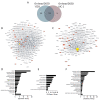
Differences between 15D3 and UIC2 IP enrichments. (A) Overlap of protein identifications between the two mAbs. (B,C) Protein interaction networks of Pgp enriched by 15D3 (B) and UIC2 (C) mAbs. Interactions were gathered and depicted using the STRING database employing all interaction sources [71] and Cytoscape software accordingly. Only proteins with at least 10 SPCs were considered. Pgp is labeled in yellow, its well-known protein partners and proteins related to MDR are highlighted in red and neighbors of these highlighted nodes are pale grey. The nodes’ size is proportional to the individual proteins’ SPCs. SPCs were normalized to all peptide-spectrum matches (PSMs) in the given analysis. Nodes of the large and small ribosome subunits and components of the chaperonin-containing T-complex were grouped together to reduce the complexity of the network. Pgp was approximately 7 times more abundant based on SPCs using UIC2 for the IP and pulled down 222 proteins, whereas with 15D3 IP a more diverse set of 368 proteins could be enriched, many of which were cholesterol-sensitive. These networks indicate that enriched proteins are indeed related to Pgp. (DF) Cellular component distribution of 15D3 enrichment (D), the 160 proteins uniquely identified in the 15D3 pull-down experiment (E), and proteins enriched in the UIC2 immunoprecipitation experiment (F) analyzed with the ClueGO app within Cytoscape [72].
Figure 2
Figure 2
Experimental approaches for XL-MS on Pgp. Cross-linking with lysine-reactive cross-linkers (DSSO and BS2Gd0/d4) was carried out either on living cells followed by membrane preparation, or on-bead after membrane preparation, then affinity purification. Cross-linked complexes containing Pgp were enriched via extracellular mAbs (15D3 and UIC2). Peptide generation via trypsin digestion was performed on-bead, and measurement of samples was realized via LC-MS/MS. Steps involved only in the living cell approach are indicated in pink, and the on-bead experimental steps are brown, while steps applied in both approaches are indicated in black.
Figure 3
Figure 3
The binding of the (A) 15D3 and (B) UIC-2 mAbs increased after PNGase F treatment applied at different concentrations on living cells as measured via flow cytometry. N-glycosyl groups sterically affect the docking of both antibodies to their binding site. (C) Western blot analysis indicates a small shift in the electrophoretic mobility of the Pgp protein after deglycosylation.
Figure 4
Figure 4
Overlap of unique mono-links on P-glycoprotein in different cross-linking setups. Each type of sample preparation was performed employing UIC2 and 15D3 monoclonal antibodies. Out of 34 unique mono-links, 6 (17.6%) were found in all sample preparation approaches; these are the ones that are most readily solvent accessible. In total, 16 (8 + 6 + 2, 47%) of the mono-links were identified at least with two different sample preparation modes which suggests that, even with distinct methods, it is possible to identify a similar set of modifications. Mono-links detected via each experimental approach visualized on Pgp’s primary structure using Protter 1.0 and on Pgp’s tertiary 6qex PDB structure can be found in Figures S1 and S2 accordingly.
Figure 8
Figure 8
ClusPro 2.0 prediction of docking (A) 15D3 mAb to Pgp (6qex); the inlet indicates binding to ECL1, ECL5 and ECL6, partially overlapping with the epitopes of (B) UIC2 (ECL1, ECL3, and ECL4), visualized by means of the UIC2-associated, 6qex PDB structure. ECLs of Pgp are highlighted in pink. NHS cross-linkers could potentially target K748 on ECL4 and K967 on ECL6; however, we did not see any modifications in these lysine residues, probably due to the vicinity of the membrane and the antibodies embedding the lysine side chains.

References

    1. Juliano R., Ling V. A Surface Glycoprotein Modulating Drug Permeability in Chinese Hamster Ovary Cell Mutants. Biochim. Biophys. Acta (BBA) -Biomembr. 1976;455:152–162. doi: 10.1016/0005-2736(76)90160-7. - DOI - PubMed
    1. Bonito C.A., Ferreira R.J., Ferreira M.J.U., Gillet J.P., Cordeiro M.N.D.S., dos Santos D.J.V.A. Theoretical Insights on Helix Repacking as the Origin of P-Glycoprotein Promiscuity. Sci. Rep. 2020;10:9823. doi: 10.1038/s41598-020-66587-5. - DOI - PMC - PubMed
    1. Rosenberg M.F., Callaghan R., Modok S., Higgins C.F., Ford R.C. Three-Dimensional Structure of P-Glycoprotein: The Transmembrane Regions Adopt an Asymmetric Configuration in the Nucleotide-Bound State. J. Biol. Chem. 2005;280:2857–2862. doi: 10.1074/jbc.M410296200. - DOI - PubMed
    1. Kopcho N., Chang G., Komives E.A. Dynamics of ABC Transporter P-Glycoprotein in Three Conformational States. Sci. Rep. 2019;9:15092. doi: 10.1038/s41598-019-50578-2. - DOI - PMC - PubMed
    1. Jiarong Li M., Guttman M., Atkins W.M. Conformational Dynamics of P-Glycoprotein in Lipid Nanodiscs and Detergent Micelles Reveal Complex Motions on a Wide Time Scale. J. Biol. Chem. 2018;293:6297–6307. doi: 10.1074/jbc.RA118.002190. - DOI - PMC - PubMed

MeSH terms

Substances

LinkOut - more resources