. 2023 Jun 26;24(13):10642.
doi: 10.3390/ijms241310642.

3K3A-Activated Protein C Inhibits Choroidal Neovascularization Growth and Leakage and Reduces NLRP3 Inflammasome, IL-1β, and Inflammatory Cell Accumulation in the Retina

Affiliations

3K3A-Activated Protein C Inhibits Choroidal Neovascularization Growth and Leakage and Reduces NLRP3 Inflammasome, IL-1β, and Inflammatory Cell Accumulation in the Retina

Yehonatan Weinberger et al. Int J Mol Sci. .

Abstract

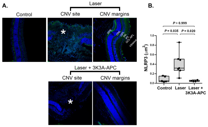
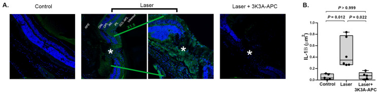
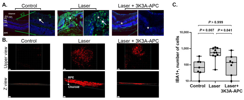
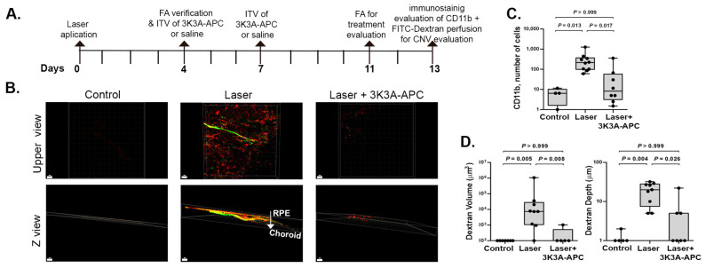
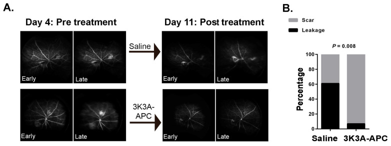
3K3A-Activated Protein C (APC) is a recombinant variant of the physiological anticoagulant APC with cytoprotective properties and reduced bleeding risks. We studied the potential use of 3K3A-APC as a multi-target therapeutic option for choroidal neovascularization (CNV), a common cause of vision loss in age-related macular degeneration. CNV was induced by laser photocoagulation in a murine model, and 3K3A-APC was intravitreally injected. The impact of 3K3A-APC treatment on myeloid and microglia cell activation and recruitment and on NLRP3 inflammasome, IL-1β, and VEGF levels was assessed using cryosection, retinal flat-mount immunohistochemistry and vascular imaging. Additionally, we evaluated the use of fluorescein angiography as a surrogate marker for in vivo evaluation of the efficacy of 3K3A-APC treatment against leaking CNV lesions. Our results demonstrated that 3K3A-APC treatment significantly reduced the accumulation and activation of myeloid cells and microglia in the CNV area and decreased the NLRP3 and IL-1β levels at the CNV site and the surrounding retina. Furthermore, 3K3A-APC treatment resulted in leakage regression and CNV growth suppression. These findings indicate that the anti-inflammatory activities of 3K3A-APC contribute to CNV inhibition. Our study suggests the potential use of 3K3A-APC as a novel multi-target treatment for CNV.

Keywords: activated protein C; choroidal neovascularization; inflammation NLRP3; microglia.


Conflict of interest statement

The authors declare no conflict of interest.

Figures

Figure 1
Figure 1
Representative figures of retinal cryosections immunostained with NLRP3 antibodies are shown in panel (A). Blue represents cell nuclei; green represents NLRP3 (scale bar = 100 µm). In control eyes, without any intervention, minimal NLRP3 expression was detected, mostly restricted to the outer part of the retina, reflecting the constitutive expression of NLRP3 inflammasome in the retina (left panel). In the eyes subjected to lasers and injected with saline, NLRP3 staining was elevated at the CNV site (marked with an asterisk) and extended to the lesion margins (Laser panels). Treatment with 3K3A-APC resulted in significant attenuation of NLRP3 staining, with minimal staining observed at the CNV site (marked with an asterisk) and the surrounding retina (Laser + 3K3A-APC panels). Quantitative analysis of the NLRP3 area (µm2) confirmed the statistical significance of these findings, as illustrated in panel (B). Positive staining areas were calculated using values from 4 microscopic fields (2 fields/slides × 2 slides/animal), which were averaged and used as raw data for further analysis. Results are presented as box-and-whiskers plots. The boxes span the 25th to the 75th percentile, the line inside each box denotes the median, and the whiskers span the lowest to the highest observations. Comparisons were performed with the Kruskal–Wallis test followed by Dunn’s post hoc test (n = 5–6 per group). CNV—Choroidal neovascularization; GCL—ganglion cell layer; INL—inner nuclear layer; IPL—inner plexiform layer; ITV—intravitreal; NFL—nerve fiber layer; ONL—outer nuclear layer; OPL—outer plexiform layer; RPE—retinal pigment epithelium; *—lesion area. NLRP3—NLR family pyrin domain containing 3.
Figure 2
Figure 2
Representative figures of retinal cryosections immunostained with IL-1β antibodies are shown in panel (A). Blue represents cell nuclei; green represents NLRP3 (scale bar = 100 µm). In control eyes without any intervention, minimal IL-1β expression was detected (left panel). IL-1β levels increased significantly after CNV induction, observed at the CNV site (marked with an asterisk) and extended to the lesion margins (middle panels). Treatment with 3K3A-APC significantly attenuated IL-1β staining, with minimal staining observed at the CNV site (marked with an asterisk) and the surrounding retina (right panel). Quantitative analysis of IL-1β area (µm2) confirmed the statistical significance of these findings, as illustrated in panel (B). Positive staining areas were calculated using values from 4 microscopic fields (2 fields/slides × 2 slides/animal), which were averaged and used as raw data for further analysis. Results are presented as box-and-whiskers plots. The boxes span the 25th to the 75th percentile, the line inside each box denotes the median, and the whiskers span the lowest to the highest observations. Comparisons were performed with the Kruskal–Wallis test followed by Dunn’s post hoc test (n = 5–6 per group). CNV—Choroidal neovascularization; GCL—ganglion cell layer; INL—inner nuclear layer; IPL—inner plexiform layer; ITV—intravitreal; NFL—nerve fiber layer; ONL—outer nuclear layer; OPL—outer plexiform layer; RPE—retinal pigment epithelium; *—lesion area. NLRP3—NLR family pyrin domain containing 3.
Figure 3
Figure 3
Representative images of retinal cryosections immunostained with IBA1 antibodies, a specific marker for microglia (green), and DAPI (blue) as a nuclei marker, are shown in panel (A). The right panel of each group represents a higher magnification (scale bar = 50 µm) of the left panel (scale bar = 100 µm). Control eyes without any intervention showed scant and ramified (non-active, marked by an arrow) IBA1+ cells, mainly in the inner retinal layers (left panel). In the untreated eyes with CNV, a significant accumulation of amoeboid, active (marked with by an arrow) microglial cells was observed in the CNV area and the surrounding retina (middle panel). However, 3K3A-APC treatment dramatically reduced microglial accumulation (right panel). IBA1+ cells and their response to 3K3A-APC treatment were further assessed in RPE-choroid flat-mounts. Intravitreal injections of either 3K3A-APC or saline were administered one hour after CNV induction. Panel (B) displays representative upper-view (upper panels) and depth Z-view (lower panels) color images of RPE-choroid flat-mounts stained with IBA1 antibodies (red) and scanned from the RPE into the choroid. Minimal staining of IBA1+ cells was detected in control eyes not subjected to laser applications (control panels). However, deeper and more extensive penetration of IBA1+ cells was observed from the RPE surface throughout the depth of the choroid in eyes exposed to lasers and treated with saline (laser panels). Treatment with 3K3A-APC significantly reduced the number and penetration of IBA1+ cells into CNV sites, and most of them were located at the edge of the RPE and not deeper into the choroid, as shown by the Z view (laser 3K3A-APC panels). Quantitative analysis of IBA1+ cells area (µm2) confirmed the statistical significance of these findings (panel (C)). Results are presented as box-and-whiskers plots. The boxes span the 25th to the 75th percentile, the line inside each box denotes the median, and the whiskers span the lowest to the highest observations. Comparisons were performed with the Kruskal–Wallis test followed by Dunn’s post hoc test (n = 7–10 per group). CNV—Choroidal neovascularization; INL—inner nuclear layer; IPL—inner plexiform layer; NFL—nerve fiber layer; ONL—outer nuclear layer; OPL—outer plexiform layer. IBA1—ionized calcium-binding adaptor molecule 1; RPE—retinal pigment epithelium; *—lesion area.
Figure 4
Figure 4
The timeline of the experiment conducted to evaluate the efficacy of 3K3A-APC treatment on myeloid cell accumulation and CNV suppression is presented in panel (A). RPE-choroid specimens were positioned with the RPE layer facing upwards and the choroid resting on the slide. Retinal blood vessels were stained using fluorescein isothiocyanate (FITC)-dextran perfusion (green), and myeloid cells were stained using anti-CD11b antibody (red), and scanned using confocal microscopy from the RPE into the choroid. Panel (B) displays representative upper-view (upper panels) and depth Z-view (lower panels) color images. Panel (C): Quantification of the total number of CD11b+ cells within the RPE-choroid specimens. 3K3A-APC treatment significantly reduces the total CD11b+ cells in the CNV area. Panel (D): Quantification of CNV volume (µm3) and penetration depth of blood vessels (µm) indicates that 3K3A-APC treatment suppresses CNV growth and reduces penetration. Results are presented as box-and-whiskers plots. The boxes span the 25th to the 75th percentile, the line inside each box denotes the median, and the whiskers span the lowest to the highest observations. Comparisons were performed with the Kruskal–Wallis test followed by Dunn’s post hoc test (n = 5–9 eyes per group). CNV—Choroidal neovascularization; FA—fluorescein angiography; RPE—retinal pigment epithelium.
Figure 5
Figure 5
Pre-treatment fluorescein angiography (FA) was conducted on day 4 post-laser to confirm the presence of leakage from CNV. Mice with confirmed leakage were then treated with 3K3A-APC or saline, and additional FA evaluated treatment efficacy on day 11 post-laser. Panel (A) shows representative dynamic FA images of the same eye, taken on days 4 and 11, which masked retina specialists used to evaluate leakage from CNV. A quantitative assessment of leaking lesions, comparing the percentage of leaking lesions in 3K3A-APC or vehicle-treated eyes, performed on day 11, is presented in panel (B). To account for 2 lesions per mouse, the data were analyzed by using a generalized estimating equation, with the lesion status (“leaking” or “non-leaking”) as a binary outcome and 3K3A-APC treatment (“no” or “yes”) as a between-subjects factor. The resulting p-value is presented in the graph. CNV—Choroidal neovascularization.

Similar articles

Cited by

References

    1. Pugazhendhi A., Hubbell M., Jairam P., Ambati B. Neovascular Macular Degeneration: A Review of Etiology, Risk Factors, and Recent Advances in Research and Therapy. Int. J. Mol. Sci. 2021;22:1170. doi: 10.3390/ijms22031170. - DOI - PMC - PubMed
    1. Fleckenstein M., Keenan T.D.L., Guymer R.H., Chakravarthy U., Schmitz-Valckenberg S., Klaver C.C., Wong W.T., Chew E.Y. Age-Related Macular Degeneration. Nat. Rev. Dis. Prim. 2021;7:31. doi: 10.1038/s41572-021-00265-2. - DOI - PubMed
    1. Wong J.H.C., Ma J.Y.W., Jobling A.I., Brandli A., Greferath U., Fletcher E.L., Vessey K.A. Exploring the Pathogenesis of Age-Related Macular Degeneration: A Review of the Interplay between Retinal Pigment Epithelium Dysfunction and the Innate Immune System. Front. Neurosci. 2022;16:1871. doi: 10.3389/fnins.2022.1009599. - DOI - PMC - PubMed
    1. Apte R.S., Chen D.S., Ferrara N. VEGF in Signaling and Disease: Beyond Discovery and Development. Cell. 2019;176:1248–1264. doi: 10.1016/j.cell.2019.01.021. - DOI - PMC - PubMed
    1. Mettu P.S., Allingham M.J., Cousins S.W. Incomplete Response to Anti-VEGF Therapy in Neovascular AMD: Exploring Disease Mechanisms and Therapeutic Opportunities. Prog. Retin. Eye Res. 2021;82:100906. doi: 10.1016/j.preteyeres.2020.100906. - DOI - PMC - PubMed

Substances

LinkOut - more resources