. 2023 Jun 26;24(13):10662.
doi: 10.3390/ijms241310662.

Involvement in Fertilization and Expression of Gamete Ubiquitin-Activating Enzymes UBA1 and UBA6 in the Ascidian Halocynthia roretzi

Affiliations

Involvement in Fertilization and Expression of Gamete Ubiquitin-Activating Enzymes UBA1 and UBA6 in the Ascidian Halocynthia roretzi

Hitoshi Sawada et al. Int J Mol Sci. .

Abstract

The extracellular ubiquitin-proteasome system is involved in sperm binding to and/or penetration of the vitelline coat (VC), a proteinaceous egg coat, during fertilization of the ascidian (Urochordata) Halocynthia roretzi. It is also known that the sperm receptor on the VC, HrVC70, is ubiquitinated and degraded by the sperm proteasome during the sperm penetration of the VC and that a 700-kDa ubiquitin-conjugating enzyme complex is released upon sperm activation on the VC, which is designated the "sperm reaction". However, the de novo function of ubiquitin-activating enzyme (UBA/E1) during fertilization is poorly understood. Here, we show that PYR-41, a UBA inhibitor, strongly inhibited the fertilization of H. roretzi. cDNA cloning of UBA1 and UBA6 from H. roretzi gonads was carried out, and their 3D protein structures were predicted to be very similar to those of human UBA1 and UBA6, respectively, based on AlphaFold2. These two genes were transcribed in the ovary and testis and other organs, among which the expression of both was highest in the ovary. Immunocytochemistry showed that these enzymes are localized on the sperm head around a mitochondrial region and the follicle cells surrounding the VC. These results led us to propose that HrUBA1, HrUBA6, or both in the sperm head mitochondrial region and follicle cells may be involved in the ubiquitination of HrVC70, which is responsible for the fertilization of H. roretzi.

Keywords: E1; FAT10; UBA; ascidian; fertilization; ubiquitin; ubiquitin-activating enzyme.


Conflict of interest statement

The authors declare no conflict of interest.

Figures

Figure 1
Figure 1
Effect of the Ub-activating enzyme inhibitor PYR-41 on fertilization of H. roretzi. Orange line, the effect of PYR-41; blue line, the effect of DMSO as a vehicle (Control). The 10 µM inhibitor contained 0.03% DMSO, while the 20 µM inhibitor contained 0.06% DMSO. Error bar, SD (n = 3). The fertilization rate was determined based on the expansion of the perivitelline space as described previously [16].
Figure 2
Figure 2
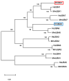
Phylogenetic tree of amino acid sequences of UBA1, UBA2, UBA3, UBA4, UBA5, UBA6, and UBA 7 from humans and mice in addition to HrUBA1 (red box) and HrUBA6 (blue box). Amino acid sequences of UBA1 (E1) and UBA6 (E1L2) from humans, mice, and the ascidian H. roretzi were aligned by Clustal Omega, and the phylogenetic tree was illustrated as described in the Section 4.
Figure 3
Figure 3
Structural visualization of HrUBA1 and HrUBA6. (A) AlphaFold2 structure prediction of HrUBA1 (left) and HrUBA6 (right). Models of HrUBA1 and HrUBA6 were generated from the local copy of Alphafold2 [30] and visualized using PyMOL by Schrödinger (https://pymol.org/2/ (accessed on 15 May 2023)) (B) HrUBA1 and HrUBA6 comprise the inactive and active adenylation domain (IAD and AAD), the first and second catalytic cysteine half domain (FCCH and SCCH), the 3-helix-bundle (3HB) and the ubiquitin fold domain (UFD) [28,29].
Figure 4
Figure 4
Expression of HrUBA1 and HrUBA6 mRNAs in respective organs by real-time PCR. cDNAs were prepared from the testis, ovary, gill, intestine, and muscles, and real-time PCR was carried out using respective cDNA preparation as a template and specific primers to HrUBA1 (A) and HrUBA6 (B). Transcription levels of HrUBA1 and HrUBA6 were normalized to the expression level of HrEF-1α. Respective columns indicate the relative expression, assuming the value in the testis as 1.0. Error bar, SD.
Figure 5
Figure 5
Localization of ascidian unfertilized eggs HrUBA1 and HrUBA6. Immunocytochemistry by using anti-HrUBA1 (A) or anti-HrUBA6 (B) antibody (upper panels) and control rabbit IgG (lower panels). Bars indicate 50 µm. PS, perivitelline space; F, follicle cells. Actin was stained with phalloidin. Follicle cells (and VC) of unfertilized eggs were stained (arrow).
Figure 6
Figure 6
Localization of HrUBA1 and HrUBA6 in sperm. Immunocytochemistry of ascidian sperm with anti-HrUBA1 (A) or anti-HrUBA6 (B) antibody (upper panels) and rabbit IgG (lower panels). Inset is an enlarged view of the area indicated by the broken line box. Bars indicate 10 µm. DNA was stained with DAPI, and mitochondria were stained with MitoTracker. The sperm mitochondrial region was stained.
Figure 7
Figure 7
Working hypothesis for localization and role in fertilization of ubiquitin-activating enzyme E1 (UBA1/6) in the ascidian Halocynthia roretzi.

Similar articles

References

    1. Hershko A., Ciechanover A. The ubiquitin system. Annu. Rev. Biochem. 1998;67:425–479. doi: 10.1146/annurev.biochem.67.1.425. - DOI - PubMed
    1. Hochstrasser M. Ubiquitin, proteasomes, and the regulation of intracellular protein degradation. Curr. Opin. Cell Biol. 1995;2:215–223. doi: 10.1016/0955-0674(95)80031-X. - DOI - PubMed
    1. Tanaka K. Proteasomes: Structure and biology. J. Biochem. 1998;123:195–204. doi: 10.1093/oxfordjournals.jbchem.a021922. - DOI - PubMed
    1. Schwartz A.L., Ciechanover A. The ubiquitin-proteasome pathway and pathogenesis of human diseases. Annu. Rev. Biochem. 1999;50:57–74. - PubMed
    1. Viestra R.D. The expanding universe of ubiquitin and ubiquitin-like modifiers. Plant Physiol. 2012;160:2–14. doi: 10.1104/pp.112.200667. - DOI - PMC - PubMed