

Clinical and Molecular Aspects of C2orf71/PCARE in Retinal Diseases
- PMID: 37445847
- PMCID: PMC10341768
- DOI: 10.3390/ijms241310670
Clinical and Molecular Aspects of C2orf71/PCARE in Retinal Diseases
Abstract
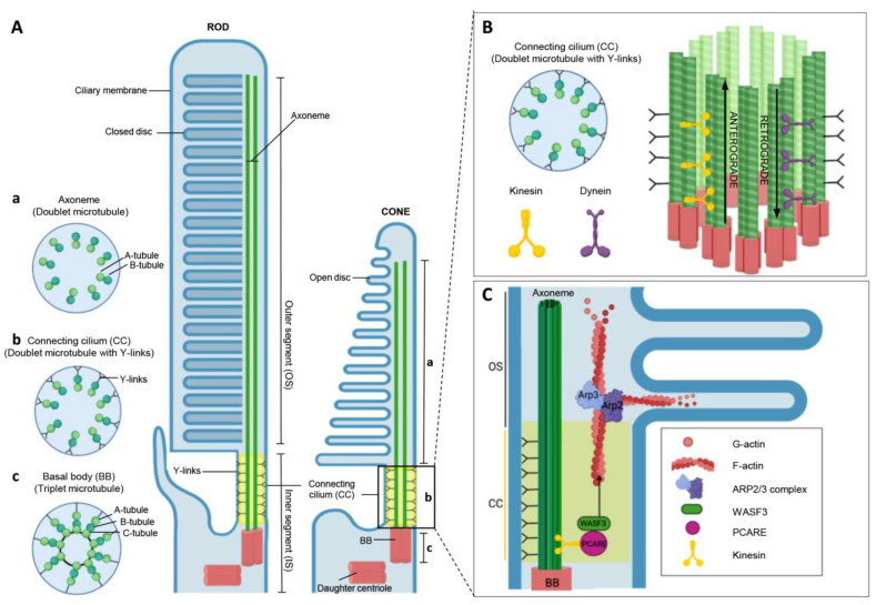
Mutations in the photoreceptor-specific C2orf71 gene (also known as photoreceptor cilium actin regulator protein PCARE) cause autosomal recessive retinitis pigmentosa type 54 and cone-rod dystrophy. No treatments are available for patients with C2orf71 retinal ciliopathies exhibiting a severe clinical phenotype. Our understanding of the disease process and the role of PCARE in the healthy retina significantly limits our capacity to transfer recent technical developments into viable therapy choices. This study summarizes the current understanding of C2orf71-related retinal diseases, including their clinical manifestations and an unclear genotype-phenotype correlation. It discusses molecular and functional studies on the photoreceptor-specific ciliary PCARE, focusing on the photoreceptor cell and its ciliary axoneme. It is proposed that PCARE is an actin-associated protein that interacts with WASF3 to regulate the actin-driven expansion of the ciliary membrane during the development of a new outer segment disk in photoreceptor cells. This review also introduces various cellular and animal models used to model these diseases and provides an overview of potential treatments.
Keywords: C2orf71 gene; PCARE; RPE; cilia; ciliopathies; cone-rod dystrophy; outer segment; photoreceptors; retina; retinitis pigmentosa.
Conflict of interest statement
The authors declare no conflict of interest.
Figures
Similar articles
-
PCARE and WASF3 regulate ciliary F-actin assembly that is required for the initiation of photoreceptor outer segment disk formation.Proc Natl Acad Sci U S A. 2020 May 5;117(18):9922-9931. doi: 10.1073/pnas.1903125117. Epub 2020 Apr 20. Proc Natl Acad Sci U S A. 2020. PMID: 32312818 Free PMC article.
-
PCARE requires coiled coil, RP62 kinase-binding and EVH1 domain-binding motifs for ciliary expansion.Hum Mol Genet. 2022 Aug 17;31(15):2560-2570. doi: 10.1093/hmg/ddac057. Hum Mol Genet. 2022. PMID: 35253837 Free PMC article.
-
C2orf71a/pcare1 is important for photoreceptor outer segment morphogenesis and visual function in zebrafish.Sci Rep. 2018 Jun 26;8(1):9675. doi: 10.1038/s41598-018-27928-7. Sci Rep. 2018. PMID: 29946172 Free PMC article.
-
Photoreceptor actin dysregulation in syndromic and non-syndromic retinitis pigmentosa.Biochem Soc Trans. 2018 Dec 17;46(6):1463-1473. doi: 10.1042/BST20180138. Epub 2018 Nov 21. Biochem Soc Trans. 2018. PMID: 30464047 Review.
-
Non-syndromic retinal ciliopathies: translating gene discovery into therapy.Hum Mol Genet. 2012 Oct 15;21(R1):R111-24. doi: 10.1093/hmg/dds298. Epub 2012 Jul 26. Hum Mol Genet. 2012. PMID: 22843501 Review.
Cited by
-
PCARE-Associated Retinopathy - Genetics, Clinical Characteristics, and Natural History.Invest Ophthalmol Vis Sci. 2025 Apr 1;66(4):61. doi: 10.1167/iovs.66.4.61. Invest Ophthalmol Vis Sci. 2025. PMID: 40261664 Free PMC article.
-
Mutational Profile and Retinal Phenotypes of PCARE-Related Cone-Rod Dystrophies in a Mexican Cohort.J Ophthalmol. 2024 Mar 4;2024:4003914. doi: 10.1155/2024/4003914. eCollection 2024. J Ophthalmol. 2024. PMID: 38468717 Free PMC article.
-
Posterior Polar Annular Choroidal Dystrophy: Genetic Insights and Differential Diagnosis in Inherited Retinal Diseases.Curr Issues Mol Biol. 2024 Feb 5;46(2):1383-1397. doi: 10.3390/cimb46020089. Curr Issues Mol Biol. 2024. PMID: 38392207 Free PMC article. Review.
-
Special Issue "The Molecular and Cellular Pathophysiologic Mechanisms Underlying Ocular Diseases and Emerging Therapies".Int J Mol Sci. 2024 Feb 18;25(4):2405. doi: 10.3390/ijms25042405. Int J Mol Sci. 2024. PMID: 38397080 Free PMC article.
References
-
- Nishimura D.Y., Baye L.M., Perveen R., Searby C.C., Avila-Fernandez A., Pereiro I., Ayuso C., Valverde D., Bishop P.N., Manson F.D.C., et al. Discovery and Functional Analysis of a Retinitis Pigmentosa Gene, C2ORF71. Am. J. Hum. Genet. 2010;86:686–695. doi: 10.1016/j.ajhg.2010.03.005. - DOI - PMC - PubMed
-
- Collin R.W.J., Safieh C., Littink K.W., Shalev S.A., Garzozi H.J., Rizel L., Abbasi A.H., Cremers F.P.M., den Hollander A.I., Klevering B.J., et al. Mutations in C2ORF71 Cause Autosomal-Recessive Retinitis Pigmentosa. Am. J. Hum. Genet. 2010;86:783–788. doi: 10.1016/j.ajhg.2010.03.016. - DOI - PMC - PubMed
-
- Mahabadi N., Khalili Y. Al Neuroanatomy, Retina. StatPearls; Tampa, FL, USA: 2022.
Publication types
MeSH terms
Substances
Grants and funding
LinkOut - more resources
Full Text Sources
