. 2023 Jun 27;24(13):10701.
doi: 10.3390/ijms241310701.

Skin Wound following Irradiation Aggravates Radiation-Induced Brain Injury in a Mouse Model

Affiliations

Skin Wound following Irradiation Aggravates Radiation-Induced Brain Injury in a Mouse Model

Mang Xiao et al. Int J Mol Sci. .

Abstract

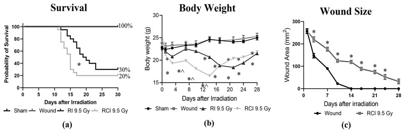
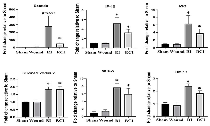
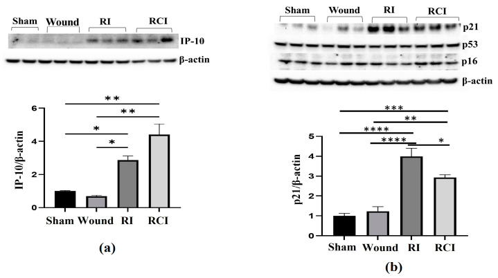
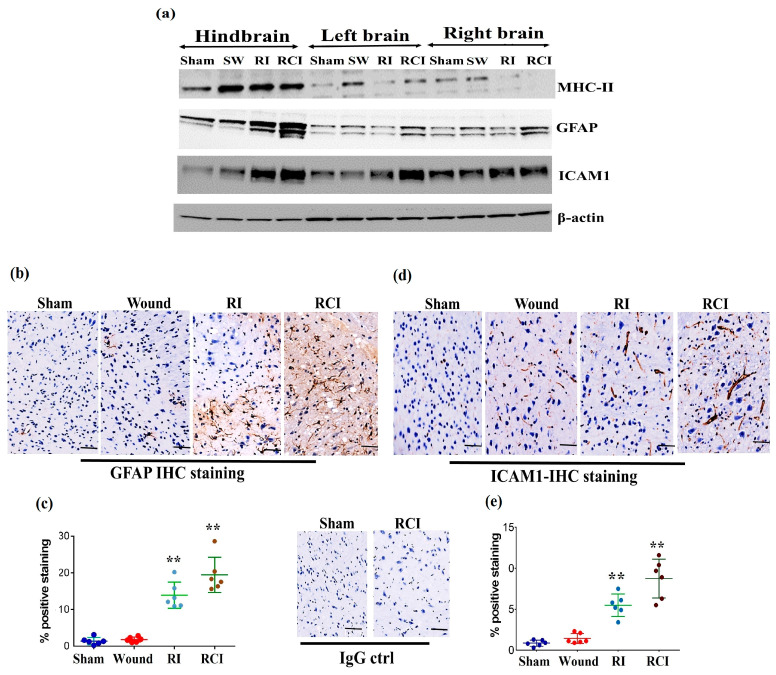
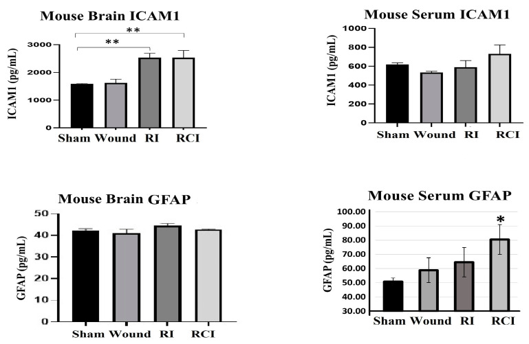
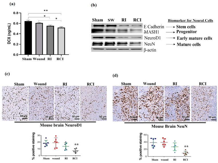
Radiation injury- and radiation combined with skin injury-induced inflammatory responses in the mouse brain were evaluated in this study. Female B6D2F1/J mice were subjected to a sham, a skin wound (SW), 9.5 Gy 60Co total-body gamma irradiation (RI), or 9.5 Gy RI combined with a skin puncture wound (RCI). Survival, body weight, and wound healing were tracked for 30 days, and mouse brain samples were collected on day 30 after SW, RI, RCI, and the sham control. Our results showed that RCI caused more severe animal death and body weight loss compared with RI, and skin wound healing was significantly delayed by RCI compared to SW. RCI and RI increased the chemokines Eotaxin, IP-10, MIG, 6Ckine/Exodus2, MCP-5, and TIMP-1 in the brain compared to SW and the sham control mice, and the Western blot results showed that IP-10 and p21 were significantly upregulated in brain cells post-RI or -RCI. RI and RCI activated both astrocytes and endothelial cells in the mouse brain, subsequently inducing blood-brain barrier (BBB) leakage, as shown by the increased ICAM1 and GFAP proteins in the brain and GFAP in the serum. The Doublecortin (DCX) protein, the "gold standard" for measuring neurogenesis, was significantly downregulated by RI and RCI compared with the sham group. Furthermore, RI and RCI decreased the expression of the neural stem cell marker E-cadherin, the intermediate progenitor marker MASH1, the immature neuron cell marker NeuroD1, and the mature neuron cell marker NeuN, indicating neural cell damage in all development stages after RI and RCI. Immunohistochemistry (IHC) staining further confirmed the significant loss of neural cells in RCI. Our data demonstrated that RI and RCI induced brain injury through inflammatory pathways, and RCI exacerbated neural cell damage more than RI.

Keywords: blood–brain barrier leakage; brain injury; inflammation; neural cell damage; radiation; radiation combined injury; skin wound.


Conflict of interest statement

The authors declare no conflict of interest. The funders had no role in the design of the study; in the collection, analysis, or interpretation of the data; in the writing of the manuscript; or in the decision to publish the results.

Figures

Figure 1
Figure 1
Effects of RI and RCI on 30-day lethality, body weight loss, and wound closure delay. (a) Thirty-day survival rate in sham-irradiated, skin wounded, 9.5 Gy RI, and 9.5 Gy RCI. RI and RCI resulted in reduction of survival to 30% and 20%, respectively. SW did not cause animal death. * p < 0.05 for RI vs. RCI on day 16 post-irradiation via Log-rank (Mantel–Cox) test. (b) RI and RCI resulted in significant body weight loss, and the RCI group lost more body weight than the RI group. * p < 0.05 vs. sham and skin wound; ^ p < 0.05, RI vs. RCI via two-way ANOVA with Tukey’s multiple comparisons tests. (c) RCI delayed wound closure compared with skin wound. * p < 0.05 via two-way ANOVA with Tukey’s multiple comparisons tests. N = 20/group in all groups.
Figure 2
Figure 2
Proinflammatory chemokine increase in mouse brains after skin wound, RI, and RCI. Brain tissues were collected from sham-irradiated, SW, RI, and RCI mice 30 days after TBI. The collected brain tissue lysates (supernatants) were subjected to a cytokine antibody array analysis (with a total of 44 antibodies for cytokines and chemokines) by a company (Eve Technologies, Calgary, AB, Canada). Six chemokines (Eotaxin, IP-10, MIG, 6Ckine, MCP-5, and TIMP-1) were markedly increased in RI and/or RCI groups of mouse brains compared with samples from sham control mouse brains (N = 4–5/group). Data are presented as means ± SEM. * p < 0.05 vs. sham-irradiated control group. Wound: skin wound; RI: 9.5 Gy; RCI: 9.5 Gy + SW.
Figure 3
Figure 3
RI and RCI upregulate IP-10 and p21 expression in mouse brain. Mouse brain tissues were collected 30 days after sham-irradiated control, skin wound, RI, and RCI. (a) IP-10, (b) p16, p21, and p53 were evaluated using an immunoblotting assay. Ratios of IP-10/β-actin and p21/β-actin are shown in histogram panels. Data are presented as means ± SEM. * p < 0.05, ** p < 0.01, *** p< 0.002, **** p< 0.001 vs. sham-irradiated control group. Western blot images are presented from one of two independent experiments (N = 4–5/group). Wound: skin wound; RI: 9.5 Gy; RCI: 9.5 Gy + SW.
Figure 4
Figure 4
RI and RCI induce microglial, endothelial cell, and astrocyte activation. (a) Mouse brain tissues were collected from left brain, right brain and hindbrain 30 days after TBI. The microglial activation marker MHC-II, the astrocyte activation marker GFAP, and the neuro-endothelial cell activation marker ICAM1 were examined via immunoblotting. Data are presented from one of two independent experiments. (be) Immunohistochemistry (IHC) staining of brains with anti-GFAP and anti-ICAM1 antibodies. Mouse brain specimens from sham-irradiated control, SW, RI, and RCI were stained with (b) anti-GFAP or (d) anti-ICAM1; scale bar = 25 µm. Brown color indicates positive staining and IgG control antibodies show negative staining. Quantification of GFAP IHC staining is shown in (c) and ICAM1 IHC staining is shown in (e). Three animals/group and two slides/animal. Data are presented as means ± SD. ** p < 0.01 vs. sham control group. Note that RCI group has the highest % of GFAP- and ICAM1-positive cells, with an enlarged cell size. SW or Wound: skin wound; RI: 9.5 Gy; RCI: 9.5 Gy + SW.
Figure 5
Figure 5
RI and RCI induce mouse blood–brain barrier (BBB) leakage. Levels of ICAM1 and GFAP in mouse brain tissue and serum from sham-irradiated control, skin wound, RI, and RCI mice were measured via ELISA (N = 4–5/group). Data are presented as means ± SD. * p< 0.05; ** p < 0.01 vs. sham-irradiated control group. Wound: skin wound; RI: 9.5 Gy; RCI: 9.5 Gy + SW.
Figure 6
Figure 6
RI and RCI inhibit neurogenesis and decrease neuron cell numbers. (a) Levels of DCX in mouse brain from sham-irradiated control, SW, RI, and RCI mice were measured via ELISA (N = 4–5/group). Data are presented as means ± SD. * p< 0.05; ** p < 0.01 vs. sham-irradiated control group. Wound: skin wound; RI: 9.5 Gy; RCI: 9.5 Gy + SW. (b) Neuron cell markers at different development stages were measured via immunoblotting assay with specific antibodies, including anti-E Cadherin (very early stage cell marker), anti-MASH1 (progenitor cell maker), anti- NeuroD1 (early mature neural cell maker), and anti-NeuN (mature neural cell maker). Data are presented from one of two independent experiments. Mouse brain specimens from sham-irradiated control, SW, RI, and RCI. Immunohistochemistry (IHC) staining of brains with anti-NeuroD1 (c) or anti- NeuN (d) antibodies was performed. Brown color indicates positive staining. Scale bar = 50 µm. Quantification of NeuroD1 and NeuN IHC staining ia shown in the histogram panels, respectively (3 animals/group and 2 slides/animal). Data are presented as means ± SD. **, p < 0.01 vs. sham-irradiated control group. Note that RCI group has significantly decreased % of neuron cells. SW or Wound: skin wound; RI: 9.5 Gy; RCI: 9.5 Gy + SW.

Similar articles

Cited by

References

    1. DiCarlo A.L., Hatchett R.J., Kaminski J.M., Ledney G.D., Pellmar T.C., Okunieff P., Ramakrishnan N. Medical Countermeasures for Radiation Combined Injury: Radiation with Burn, Blast, Trauma and/or Sepsis. Report of an NIAID Workshop, March 26–27, 2007. Radiat. Res. 2008;169:712–721. doi: 10.1667/RR1295.1. - DOI - PMC - PubMed
    1. Wang L., Zhai M., Lin B., Cui W., Hull L., Li X., Anderson M.N., Smith J.T., Umali M.V., Jiang S., et al. PEG-G-CSF and L-Citrulline Combination Therapy for Mitigating Skin Wound Combined Radiation Injury in a Mouse Model. Radiat. Res. 2021;196:113–127. doi: 10.1667/RADE-20-00151.1. - DOI - PMC - PubMed
    1. Wang L., Lin B., Zhai M., Cui W., Hull L., Zizzo A., Li X., Kiang J.G., Xiao M. Deteriorative Effects of Radiation Injury Combined with Skin Wounding in a Mouse Model. Toxics. 2022;10:785. doi: 10.3390/toxics10120785. - DOI - PMC - PubMed
    1. Hauer-Jensen M., Kumar K.S., Wang J., Berbee M., Fu Q., Boerma M. Intestinal Toxicity in Radiation- and Combined Injury: Significance, Mechanisms, and Countermeasures: 2008. Nova Science Publishers, Inc.; New York, NY, USA: 2008. pp. 61–100.
    1. Kiang J.G., Jiao W., Cary L.H., Mog S.R., Elliott T.B., Pellmar T.C., Ledney G.D. Wound Trauma Increases Radiation-Induced Mortality by Activation of iNOS Pathway and Elevation of Cytokine Concentrations and Bacterial Infection. Radiat. Res. 2010;173:319–332. doi: 10.1667/RR1892.1. - DOI - PMC - PubMed

Substances