. 2023 Jun 27;24(13):10720.
doi: 10.3390/ijms241310720.

I-Shaped Dimers of a Plant Chloroplast FOF1-ATP Synthase in Response to Changes in Ionic Strength

Affiliations

I-Shaped Dimers of a Plant Chloroplast FOF1-ATP Synthase in Response to Changes in Ionic Strength

Stepan D Osipov et al. Int J Mol Sci. .

Abstract

F-type ATP synthases play a key role in oxidative and photophosphorylation processes generating adenosine triphosphate (ATP) for most biochemical reactions in living organisms. In contrast to the mitochondrial FOF1-ATP synthases, those of chloroplasts are known to be mostly monomers with approx. 15% fraction of oligomers interacting presumably non-specifically in a thylakoid membrane. To shed light on the nature of this difference we studied interactions of the chloroplast ATP synthases using small-angle X-ray scattering (SAXS) method. Here, we report evidence of I-shaped dimerization of solubilized FOF1-ATP synthases from spinach chloroplasts at different ionic strengths. The structural data were obtained by SAXS and demonstrated dimerization in response to ionic strength. The best model describing SAXS data was two ATP-synthases connected through F1/F1' parts, presumably via their δ-subunits, forming "I" shape dimers. Such I-shaped dimers might possibly connect the neighboring lamellae in thylakoid stacks assuming that the FOF1 monomers comprising such dimers are embedded in parallel opposing stacked thylakoid membrane areas. If this type of dimerization exists in nature, it might be one of the pathways of inhibition of chloroplast FOF1-ATP synthase for preventing ATP hydrolysis in the dark, when ionic strength in plant chloroplasts is rising. Together with a redox switch inserted into a γ-subunit of chloroplast FOF1 and lateral oligomerization, an I-shaped dimerization might comprise a subtle regulatory process of ATP synthesis and stabilize the structure of thylakoid stacks in chloroplasts.

Keywords: FOF1-ATP synthase; chloroplasts; dimers; membrane proteins; small-angle scattering.


Conflict of interest statement

The authors declare no conflict of interest.

Figures

Figure 1
Figure 1
Purification and characterization of cFOF1: (a) Overall view of ATP synthase from spinach chloroplasts. FO part consists of abb′c14 subunits, F1 part consists of α3β3γεδ subunits and bb′ subunits form a peripheral stalk. The structure of ATP synthase from spinach chloroplasts (PDB ID: 6FKF [15]) was used for representation; (b) Characterization of the peak fractions of cFOF1 after anion exchange chromatography (AEX) by SAXS, experimental I(Q) 1D-profile showed as hollow grey stars, a model of cFOF1 (PDB ID: 6FKF) with a detergent belt was used for approximation of experimental data with χ2 = 1.15 (blue line); (c) Characterization of the AEX peak fractions of cFOF1 by SDS PAGE, colored with Coomassie; (d) Anion exchange chromatography (AEX) of cFOF1 from spinach chloroplasts, highlighted fractions were merged and taken for structural studies, the region shown with a red box is described in details in Panel (e); (e) AEX peak fractions of cFOF1. Panels (a,ce) were adapted from [12] with modifications.
Figure 2
Figure 2
SAXS characterization of cFOF1: (a) Experimental I(Q) 1D-profiles for AEX-purified and dialyzed against the same buffer with different NaCl concentrations samples of cFOF1, experimental data are shown as dots, regularized fits are shown as black lines. For clarity, SAXS data for 150, 250, 300, 350, and 450 mM NaCl were multiplied by 10, 102, 103, 104, and 105, respectively; (b) Normalized pair-distance distribution function P(r) for cFOF1 at different NaCl concentrations; (c) Maximum size of the object (Dmax) for samples of cFOF1 at different NaCl concentrations; (d) Radius of gyration (Rg) for samples of cFOF1 at different NaCl concentrations obtained from P(r); (e) Porod volume (Vp) for samples of cFOF1 at different NaCl concentrations. Gray lines show the values of Dmax, Rg, Vp for an AEX-purified sample of cFOF1 without dialysis. The values used for plots in panels (ce) are given in Table S1.
Figure 3
Figure 3
Dimerization of cFOF1 from Spinacia oleracea at 450 mM NaCl shown by SAXS: (a) Experimental I(Q) 1D-profile for cFOF1 at 450 mM NaCl (orange squares) and an approximation (blue line, χ2 = 2.36) by using a model of a mixture of cFOF1 monomers and dimers formed by FO-FO contacts (see Macromolecular docking section in Materials and Methods); (b) Experimental I(Q) 1D-profile for cFOF1 at 450 mM NaCl (the same as in panel (a), orange squares) and an approximation (blue line, χ2 = 1.20) by using a model of a mixture of cFOF1 monomers and dimers formed by F1-F1 contacts (see Macromolecular docking section in Materials and Methods); (c) Relative residues of the fit shown in panel (a); (d) Relative residues of the fit shown in panel (b). The models of monomeric and dimeric cFOF1, shown in panels (a,b), contain detergent belts obtained from the experimental SAXS data and built by the program MEMPROT. Volume fractions of monomers and dimers of cFOF1, used in the models for approximation, are shown.
Figure 4
Figure 4
Possible biological interpretation of observed cFOF1 I-shaped dimer: (a) Segmented subvolume of a thylakoid stack with connected stroma lamellae, cFOF1 are shown as yellow 12-nm spheres, cFOF1 is randomly distributed over stromal lamellae; (b) Surface rendering of a lateral cFOF1 dimer in isolated pea thylakoid membranes; (c) A model of an I-shaped dimer with F1/F1 interaction interface, presumably via δ-subunit, the distance between membrane FO parts of different cFOF1 monomers is about 30 nm; (d) Electron Tomography of Vitreous Spinach Chloroplast Sections, stacked grana and unstacked stroma thylakoid membranes are shown, the distance between neighboring stroma lamellae approximately fits the size of the I-shaped cFOF1 dimer, the stroma thylakoids, which are continuous with a grana thylakoid is shown with green arrowheads, the stroma thylakoids merged with two adjacent grana thylakoids—blue arrowheads, the region shown with a red box is described in details in Panel (e); (e) Surface representation of connections between grana (green) and stroma (purple) thylakoids, the distance between neighboring stroma lamellae is about 30 nm, the region shown with a green box is described in details in Panel (f); (f) Schematic representation of an I-shaped cFOF1 dimer in between of two neighboring stroma lamellae; (g) A part of a tomographic slice showing neighboring stroma lamellae, the distance between them approximately fits the size of an I-shaped ATP-synthase dimer, Pink lines are 30 nm size and demonstrate the possible fit of an I-shaped cFOF1 dimer between the neighbor lamellae. Presumably, ATP synthases shown with red arrows might be these I-shaped dimers. Yellow arrowheads show separate cFOF1. Panels (a,b,d,e,g) were adapted from [3] with modifications.
Figure 5
Figure 5
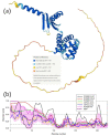
Structure and disorder in the cFOF1 δ-subunit: (a) 3D structure of the cFOF1 δ-subunit generated by AlphaFold2; (b) Per-residue intrinsic disorder predisposition of the cFOF1 δ-subunit from the spinach chloroplasts. The intrinsic disorder profile was generated using the outputs of the Rapid Intrinsic Disorder Analysis Online (RIDAO) platform [38] that aggregates the results from a number of well-known disorder predictors, such as PONDR® VLXT, PONDR® VL3, PONDR® VLS2, PONDR® FIT, and IUPred2 (Short) and IUPred2 (Long) and also produces the mean disorder profile (MDP) and corresponding error distribution (pink shadow). The outputs of the evaluation of the per-residue disorder propensity by these tools are represented as real numbers between 1 (ideal prediction of disorder) and 0 (ideal prediction of order). A threshold of 0.5 is used to identify disordered residues and regions in query proteins. Residues with the disorder scores (DS) DS < 0.15 are considered as ordered, residues with 0.15 ≤ DS < 0.25 are taken as flexible, whereas residues with 0.25 ≤ DS < 0.5 are considered as moderately disordered.

References

    1. Vlasov A.V., Osipov S.D., Bondarev N.A., Uversky V.N., Borshchevskiy V.I., Yanyushin M.F., Manukhov I.V., Rogachev A.V., Vlasova A.D., Ilyinsky N.S., et al. ATP synthase FOF1 structure, function, and structure-based drug design. Cell. Mol. Life Sci. 2022;79:179. doi: 10.1007/s00018-022-04153-0. - DOI - PMC - PubMed
    1. Kühlbrandt W. Structure and Mechanisms of F-Type ATP Synthases. Annu. Rev. Biochem. 2019;88:515–549. doi: 10.1146/annurev-biochem-013118-110903. - DOI - PubMed
    1. Daum B., Nicastro D., Austin J., McIntosh J.R., Kühlbrandt W. Arrangement of Photosystem II and ATP Synthase in Chloroplast Membranes of Spinach and Pea. Plant Cell. 2010;22:1299–1312. doi: 10.1105/tpc.109.071431. - DOI - PMC - PubMed
    1. Rexroth S., zu Tittingdorf J.M.M., Schwaßmann H.J., Krause F., Seelert H., Dencher N.A. Dimeric H+-ATP synthase in the chloroplast of Chlamydomonas reinhardtii. Biochim. Biophys. Acta Bioenerg. 2004;1658:202–211. doi: 10.1016/j.bbabio.2004.05.014. - DOI - PubMed
    1. Schwaßmann H.J., Rexroth S., Seelert H., Dencher N.A. Metabolism controls dimerization of the chloroplast FoF1ATP synthase in Chlamydomonas reinhardtii. FEBS Lett. 2007;581:1391–1396. doi: 10.1016/j.febslet.2007.02.057. - DOI - PubMed

MeSH terms

LinkOut - more resources