. 2023 Jun 27;24(13):10740.
doi: 10.3390/ijms241310740.

Extensive Bioinformatics Analyses Reveal a Phylogenetically Conserved Winged Helix (WH) Domain (Zτ) of Topoisomerase IIα, Elucidating Its Very High Affinity for Left-Handed Z-DNA and Suggesting Novel Putative Functions

Affiliations

Extensive Bioinformatics Analyses Reveal a Phylogenetically Conserved Winged Helix (WH) Domain (Zτ) of Topoisomerase IIα, Elucidating Its Very High Affinity for Left-Handed Z-DNA and Suggesting Novel Putative Functions

Martin Bartas et al. Int J Mol Sci. .

Abstract

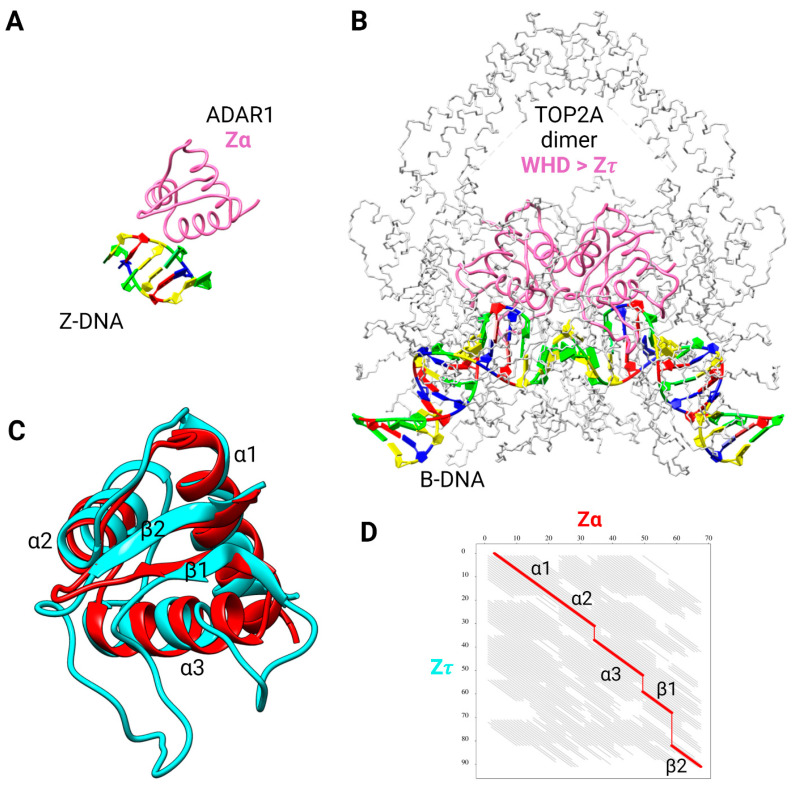
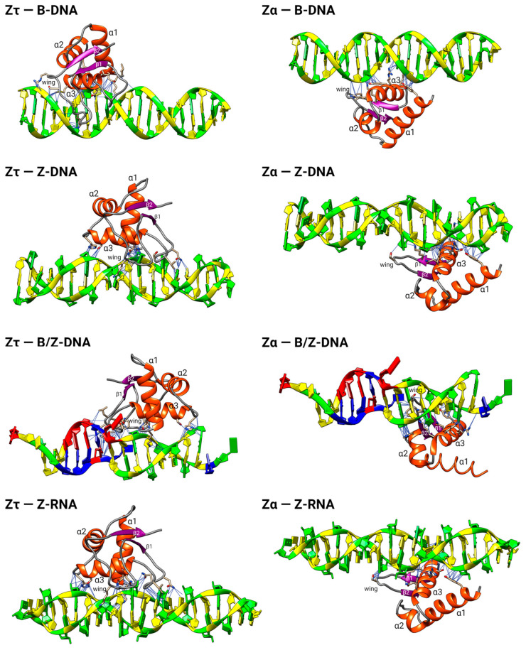
The dynamic processes operating on genomic DNA, such as gene expression and cellular division, lead inexorably to topological challenges in the form of entanglements, catenanes, knots, "bubbles", R-loops, and other outcomes of supercoiling and helical disruption. The resolution of toxic topological stress is the function attributed to DNA topoisomerases. A prominent example is the negative supercoiling (nsc) trailing processive enzymes such as DNA and RNA polymerases. The multiple equilibrium states that nscDNA can adopt by redistribution of helical twist and writhe include the left-handed double-helical conformation known as Z-DNA. Thirty years ago, one of our labs isolated a protein from Drosophila cells and embryos with a 100-fold greater affinity for Z-DNA than for B-DNA, and identified it as topoisomerase II (gene Top2, orthologous to the human UniProt proteins TOP2A and TOP2B). GTP increased the affinity and selectivity for Z-DNA even further and also led to inhibition of the isomerase enzymatic activity. An allosteric mechanism was proposed, in which topoII acts as a Z-DNA-binding protein (ZBP) to stabilize given states of topological (sub)domains and associated multiprotein complexes. We have now explored this possibility by comprehensive bioinformatic analyses of the available protein sequences of topoII representing organisms covering the whole tree of life. Multiple alignment of these sequences revealed an extremely high level of evolutionary conservation, including a winged-helix protein segment, here denoted as Zτ, constituting the putative structural homolog of Zα, the canonical Z-DNA/Z-RNA binding domain previously identified in the interferon-inducible RNA Adenosine-to-Inosine-editing deaminase, ADAR1p150. In contrast to Zα, which is separate from the protein segment responsible for catalysis, Zτ encompasses the active site tyrosine of topoII; a GTP-binding site and a GxxG sequence motif are in close proximity. Quantitative Zτ-Zα similarity comparisons and molecular docking with interaction scoring further supported the "B-Z-topoII hypothesis" and has led to an expanded mechanism for topoII function incorporating the recognition of Z-DNA segments ("Z-flipons") as an inherent and essential element. We further propose that the two Zτ domains of the topoII homodimer exhibit a single-turnover "conformase" activity on given G(ate) B-DNA segments ("Z-flipins"), inducing their transition to the left-handed Z-conformation. Inasmuch as the topoII-Z-DNA complexes are isomerase inactive, we infer that they fulfill important structural roles in key processes such as mitosis. Topoisomerases are preeminent targets of anti-cancer drug discovery, and we anticipate that detailed elucidation of their structural-functional interactions with Z-DNA and GTP will facilitate the design of novel, more potent and selective anti-cancer chemotherapeutic agents.

Keywords: GTP; Z-DNA; bioinformatics; topoII; topoisomerase IIα.


Conflict of interest statement

The authors declare no conflict of interest. The funders had no role in the design of the study; in the collection, analyses, or interpretation of data; in the writing of the manuscript; or in the decision to publish the results.

Figures

Figure 4
Figure 4
Structural comparison of Zα domain from human ADAR1p150 and putative Z𝜏 domain from human topoII. (A) Crystal structure of Zα in complex with d(CACGTG) (PDB: 3f21). (B) Cryo-EM structure of the human TOP2A DNA-binding/cleavage domain in State 1 (PDB: 6zy5). Zα or Z𝜏 domains are colored in hot pink, and DNA is colored according to NDB standards (A in red, T in blue, C in yellow, and G in green). (C) Superposition of Zα (ADAR1p150, red) and Z𝜏 (TOP2A 722–812, cyan) domains, canonical designation of helices and β-sheets is indicated. (D) Graph of FATCAT [60] chaining result intuitively showing structural similarity across all 91 aa-long alignment (thick red diagonals); three gaps are depicted using thin vertical red lines, and non-significant structural similarity is depicted by gray diagonals.
Figure 5
Figure 5
Domain composition of topoII together with putative Z-DNA-binding domains, Zτ (red), and predicted GTP-binding sites. ATP-binding sites (HATPase_c) and TOP (TOP4C) were annotated using a SMART web server [68]. (A) Known and newly identified features (Zτ, GTP-binding site, and deleterious SNPs, here depicted as Ds) in human TOP2A. (B) Zτ sequence (722–812), secondary structure, including the proposed “Z-discrimination region” (775–796, underlined residues; Table 3 below). (C) Evolutionary conservation of Zτ and GTP-binding sites in diverse eukaryotic species.
Figure 6
Figure 6
Multiple sequence alignment of TOP2 protein sequence in representative species showing very high conservation of the region Zτ constituting the Zα structural homolog (bounded by green and red triangle marks) and putative GTP-binding site. The purple asterisk mark highlights the locus critical for DNA bending. Experimentally validated ATP, Mg2+, and DNA-binding sites, together with the critical tyrosine 805 active site, are depicted as well. Columns highlighted in red and yellow show evolutionarily conserved regions/amino acid positions (primary sequence); the resulting consensus sequence is displayed in the bottom row, using criteria from MultAlin [69]: uppercase is identity, lowercase is consensus level > 0.5, ! is any-one of IV, $ is anyone of LM, % is anyone of FY, # is anyone of NDQE.
Figure 7
Figure 7
DNA Topoisomerase type IIA diversity (A) and GTP-binding (B,C). (A) Known diversity of Topoisomerase type IIA (IPR001241). According to InterPro Database, there are more than 105 protein sequences in more than 50,000 different species across the whole tree of life. (B) GTP docked to the crystal structure of human topoII (PDB: 4fm9). Only the immediate surrounding of the docked GTP molecule is shown, but it comprises both the predicted GTP-binding region (in green) and Z𝜏. The tyrosine active site of TOP2A is in red. (C) Sequence logo of the region corresponding to the putative GTP-binding site, and the GxxG/GxxxG motif(s) based on seed alignment of the DNA topoisomerase IV (PF00521) domain. The logo was produced using the Skylign tool [71].
Figure 8
Figure 8
Molecular docking for various interacting protein–DNA pairs. The left column is for Zτ from the human protein TOP2A and the right column for Zα from the human protein ADAR1p150, both docked to the indicated nucleic acid structures (B-DNA, Z-DNA, B/Z-DNA, and Z-RNA).
Figure 9
Figure 9
“B-Z-TopoII”: expanded reaction mechanism of topoII incorporating Z-DNA binding and a postulated conformase activity in addition to its canonical isomerase function. (A) Functional scheme, explained in the text. (B) Details of intermediate states (in square brackets) and outcomes of the isomerase and conformase pathways. The gray shaded area comprises the “DNA manifold” with interconversions between linear and bent conformations that depend on sequence, topological state, solution conditions, and external factors. Straight lines denote interactions between binding partners, leading to reactions (lines with arrows; in reversible reactions, a larger arrowhead indicates preferential state). Configurations of the topoII homodimer: To, free; TB; bound bent (Bb) B-DNA (G-segment); TZ; configuration bound to bent (Zb) (G-segment); TBA, TB with bound ATP; TZG, TZ with bound GTP. Adapted from Figure 1 in Ref. [78].
Figure 1
Figure 1
Comparative structures of alternative conformations of dsDNA: left, B-DNA; right, Z-DNA; center, a B-Z composite DNA with an intervening junction. Black dashed arrows indicate handedness (B-DNA, right; Z-DNA, left). Horizontal arrows indicate transitions between depicted dsDNA conformations. The figure was constructed with UCSF Chimera: B-DNA and Z-DNA are modeled structures, and B/Z-DNA is a crystal structure (PDB: 5zup). The helical pitch of B-DNA is 33 nm (10.5 bp/turn), and for Z-DNA, it is 46 nm (12 bp/turn).
Figure 2
Figure 2
Prior bioinformatic and biochemical modeling instigating the search for a Z-DNA recognition domain of topoII. (A) Representative molecular docking of human HOP2 Zα region (aa 1–74) to Z-DNA [26]. Potential key amino acid interactions are depicted by thin blue lines. (B) Example of topoII as an allosteric ZBP (left and right). It is subject to competition by molecules (middle) exerting isomerase activity on nsc B-DNA segments. The relaxation process (ellipse with arrow, long curved line) abrogates the Z-conformation (short curved line) in the designated topologically linked segments. The affinity of topoII for Z-DNA is much greater than for B-DNA and increases further in the presence of GTP (topoII*), which also inhibits isomerase function (property 12, Table 1). These binding sites are deemed to constitute potential clamps, barriers, and crosslinkers, for example, in chromatin remodeling and mitosis/meiosis. Adapted from Figure 9 of Ref. [8].
Figure 3
Figure 3
Canonical triple-gate isomerase mechanism for topoII. A double-helical DNA segment (G) binds to the topoII homodimer (upper left), is bent in the process, and is then cleaved, resulting in covalent protein (tyrosine)-DNA intermediates demarcating a double-strand break (DSB). A second “captured” DNA segment (T) traverses the DSB and is then released, while the G segment is religated, thereby restoring the integrity of the double helix. The intricate, sequential, concerted process is under allosteric control [48] mediated by ATP binding and turnover ([49], colored asterisks), divalent cations [50,51], and protein domains subject to post-translational modification, notably of the CTD [48]. The open and closed clamps of stages 1 and 3, respectively, are well depicted in a model of the tobacco enzyme (Figure 6 of Ref. [52]). Each cycle comprises a dual strand passage and thus changes the topological linking number Lk by ±2. The juxtaposition (a more appropriate term might be apposition) of the G and T segments at the crossover locus is dictated by the 3D structure of the local DNA domain, leading to numerous alternative topological outcomes [53]: resolution/simplification (relaxation, disentanglement) of plectonemic and toroidal supercoiled (+,−) substructures and reversal/formation of knots and catenanes arising during the processes of DNA transcription, replication, repair, recombination, higher-order chromosomal restructuring during mitosis and meiosis, and processing of closed circular DNA. Interference with DSB formation and resealing is highly genotoxic, and thus, steps 2 and 3 are key targets of antimicrobial and anticancer drugs [54,55,56,57,58]. Adapted from Figure 4 of Ref. [30].

Similar articles

Cited by

References

    1. Wang J.C. Untangling the Double Helix: DNA Entanglement and the Action of the DNA Topoisomerases. Cold Spring Harbor Laboratory Press; New York, NY, USA: 2009.
    1. Hanke A., Ziraldo R., Levene S.D. DNA-Topology Simplification by Topoisomerases. Molecules. 2021;26:3375. doi: 10.3390/molecules26113375. - DOI - PMC - PubMed
    1. Pommier Y., Nussenzweig A., Takeda S., Austin C. Human Topoisomerases and Their Roles in Genome Stability and Organization. Nat. Rev. Mol. Cell Biol. 2022;23:407–427. doi: 10.1038/s41580-022-00452-3. - DOI - PMC - PubMed
    1. Vidmar V., Vayssières M., Lamour V. What’s on the Other Side of the Gate: A Structural Perspective on DNA Gate Opening of Type IA and IIA DNA Topoisomerases. Int. J. Mol. Sci. 2023;24:3986. doi: 10.3390/ijms24043986. - DOI - PMC - PubMed
    1. Neidle S., Sanderson M. Principles of Nucleic Acid Structure. 2nd ed. Academic Press; Cambridge, MA, USA: 2021.

LinkOut - more resources