. 2023 Jun 28;24(13):10752.
doi: 10.3390/ijms241310752.

Does Diffusely Infiltrating Lobular Carcinoma of the Breast Arise from Epithelial-Mesenchymal Hybrid Cells?

Affiliations

Does Diffusely Infiltrating Lobular Carcinoma of the Breast Arise from Epithelial-Mesenchymal Hybrid Cells?

László Tabár et al. Int J Mol Sci. .

Abstract

Classic diffusely infiltrating lobular carcinoma has imaging features divergent from the breast cancers originating from the terminal ductal lobular units and from the major lactiferous ducts. Although the term "invasive lobular carcinoma" implies a site of origin within the breast lobular epithelium, we were unable to find evidence supporting this assumption. Exceptional excess of fibrous connective tissue and the unique cell architecture combined with the aberrant features at breast imaging suggest that this breast malignancy has not originated from cells lining the breast ducts and lobules. The only remaining relevant component of the fibroglandular tissue is the mesenchyme. The cells freshly isolated and cultured from diffusely infiltrating lobular carcinoma cases contained epithelial-mesenchymal hybrid cells with both epithelial and mesenchymal properties. The radiologic and histopathologic features of the tumours and expression of the mesenchymal stem cell positive markers CD73, CD90, and CD105 all suggest development in the direction of mesenchymal transition. These hybrid cells have tumour-initiating potential and have been shown to have poor prognosis and resistance to therapy targeted for malignancies of breast epithelial origin. Our work emphasizes the need for new approaches to the diagnosis and therapy of this highly fatal breast cancer subtype.

Keywords: biomarkers; breast neoplasms; early detection of cancer; histopathology technology; interdisciplinary communication; mammography; neoplastic stem cell; pathologists; patient care; precision oncology.


Conflict of interest statement

The authors declare no conflict of interest.

Figures

Figure A1
Figure A1
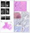
Case 2. Imaging and histopathological findings. Mediolateral (a) and craniocaudal (b) mammograms showing a normal left breast and an extensive region of architectural distortion in the upper-outer quadrant of the right breast with no associated calcifications. Handheld ultrasound shows an anechoic malignant tumour (c,d). Magnetic resonance imaging demonstrates the architectural distortion of the enhancing tumour (e). Low-power, large-format histopathology image of this 42 mm moderately differentiated, diffusely infiltrating carcinoma (f). Higher-power histopathology image shows features of a “classic infiltrating lobular carcinoma” (g). No lymphovascular, perivascular, or perineural invasions were demonstrable. The immunohistochemical staining showed strong diffuse oestrogen receptor (h) and progesterone receptor (i) positivity in 95% of the tumour cells, human epidermal growth factor receptor 2 (HER-2) negativity (j), 10% proliferation index (Ki 67) (k), and the absence of E-cadherin (l). (magnification: 10×).
Figure A2
Figure A2
(A) Case 3. Imaging and histopathological findings in the left breast. Mediolateral (a,b) and craniocaudal (c,d) mammograms. Corresponding to the palpable tumour in the upper-outer quadrant of the left breast, there is a diffuse, non-specific asymmetric density with no associated calcifications, extending from the retracted nipple to the chest wall (b,d). There is a mammographically detected, 20 mm stellate tumour mass in the upper-inner quadrant of the right breast (a,c) (see description in (B)). Magnetic resonance images (e,f) demonstrate the extensive non-mass enhancement in the left breast and the small stellate tumour mass in the right breast (see description in (B)). Handheld ultrasound shows a large anechoic malignant tumour in the left breast (g). Low-power, large-format histopathology image (h) of a portion of this histologically 75 mm moderately differentiated, diffusely infiltrating carcinoma. The histopathologic features of a “classic infiltrating lobular carcinoma” are shown on the higher-power images of the left breast specimen (i). The immunohistochemical staining showed strong diffuse oestrogen receptor (j) and progesterone receptor (k) positivity in 95% of the tumour cells, human epidermal growth factor receptor 2 (HER-2) negativity (l), 5% proliferation index (Ki 67) (m), and the absence of E-cadherin (n). (B) Case 3. Imaging and histopathological findings in the right breast. Mediolateral (a,b) and craniocaudal (c,d) mammograms. There is a mammographically detected, 18 mm stellate tumour mass in the upper-inner quadrant of the right breast (a,c). Microfocus magnification of the mammographically malignant stellate tumour (e). Handheld ultrasound of the right breast confirms the malignant appearance of this small stellate tumour (f). Magnetic resonance images of the right breast demonstrate a unifocal stellate tumour mass (g,h). Low-power, large-format histopathology image of this 18 mm invasive breast cancer (i). Intermediate (j) and high power (k) histopathology images of this moderately differentiated breast cancer, acinar adenocarcinoma of the breast, AAB. The immunohistochemical staining showed diffuse oestrogen receptor (l) and progesterone receptor (m) positivity in 90% of the tumour cells, human epidermal growth factor receptor 2 (HER-2) negativity (n), 5% proliferation index (Ki 67) (o), and strongly positive E-cadherin staining (p). (magnification: 10×).
Figure A2
Figure A2
(A) Case 3. Imaging and histopathological findings in the left breast. Mediolateral (a,b) and craniocaudal (c,d) mammograms. Corresponding to the palpable tumour in the upper-outer quadrant of the left breast, there is a diffuse, non-specific asymmetric density with no associated calcifications, extending from the retracted nipple to the chest wall (b,d). There is a mammographically detected, 20 mm stellate tumour mass in the upper-inner quadrant of the right breast (a,c) (see description in (B)). Magnetic resonance images (e,f) demonstrate the extensive non-mass enhancement in the left breast and the small stellate tumour mass in the right breast (see description in (B)). Handheld ultrasound shows a large anechoic malignant tumour in the left breast (g). Low-power, large-format histopathology image (h) of a portion of this histologically 75 mm moderately differentiated, diffusely infiltrating carcinoma. The histopathologic features of a “classic infiltrating lobular carcinoma” are shown on the higher-power images of the left breast specimen (i). The immunohistochemical staining showed strong diffuse oestrogen receptor (j) and progesterone receptor (k) positivity in 95% of the tumour cells, human epidermal growth factor receptor 2 (HER-2) negativity (l), 5% proliferation index (Ki 67) (m), and the absence of E-cadherin (n). (B) Case 3. Imaging and histopathological findings in the right breast. Mediolateral (a,b) and craniocaudal (c,d) mammograms. There is a mammographically detected, 18 mm stellate tumour mass in the upper-inner quadrant of the right breast (a,c). Microfocus magnification of the mammographically malignant stellate tumour (e). Handheld ultrasound of the right breast confirms the malignant appearance of this small stellate tumour (f). Magnetic resonance images of the right breast demonstrate a unifocal stellate tumour mass (g,h). Low-power, large-format histopathology image of this 18 mm invasive breast cancer (i). Intermediate (j) and high power (k) histopathology images of this moderately differentiated breast cancer, acinar adenocarcinoma of the breast, AAB. The immunohistochemical staining showed diffuse oestrogen receptor (l) and progesterone receptor (m) positivity in 90% of the tumour cells, human epidermal growth factor receptor 2 (HER-2) negativity (n), 5% proliferation index (Ki 67) (o), and strongly positive E-cadherin staining (p). (magnification: 10×).
Figure 1
Figure 1
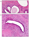
Details of large-format thin-section histopathology images of diffusely infiltrating lobular carcinoma. The invasive carcinoma surrounds the lobular acini (a) and a duct (b). The epithelial cells of both the lobular acini and the duct are normal, free of atypia, and free of evidence of lobular hyperplasia or LCIS (lobular cancer in situ), despite scrutiny using large-format sections (hematoxylin-eosin staining (H&E), 10× magnification).
Figure 2
Figure 2
Cultured cells from diffusely infiltrating breast cancer. Long-term follow-up of the cells isolated from diffusely infiltrating breast cancer in Dulbecco’s Modified Eagle Medium (DMEM) with high glucose (a) and in RPMI-1640 medium (b). The cell growth was monitored by phase-contrast microscopy (20× original magnification). Dashed lines indicate the enlarged regions of the images.
Figure 3
Figure 3
Microtumour-like phenotype of cells derived from diffusely infiltrating breast cancer showing the ability of the cultured cells to form microtumours regardless of time, culture media, and passage number. Cells were cultured in RPMI-1640 medium (20× original magnification) (a). Follow-up of a microtumour grown in Dulbecco’s Modified Eagle Medium (DMEM) (5× original magnification) (b).
Figure 4
Figure 4
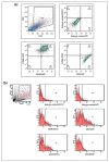
Flow cytometric analysis of the mesenchymal stem cell surface markers. Representative plots of the analysis of the mesenchymal stem cell related markers, CD34, CD73, CD90, and CD105 on the surfaces of cells originating from the three cases cultured in RPMI-1640 medium in the 4th passage (a) and on the surfaces of the freshly isolated cells (b).
Figure 5
Figure 5
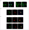
Mesenchymal-like characteristics of the cultured cells. Immunofluorescence staining for cytokeratin AE1-AE3 and smooth-muscle actin (a), CD73-cytokeratin 5/8 (b), and CD73-OKT4 and CD73-Nanog (c) in cells cultured from diffusely infiltrating breast cancer in RPMI-1640 medium (n = 3). Nuclei were visualized by 4′,6-diamidino-2-phenylindole (DAPI) staining (40× original magnification with immersion oil; Zeiss Axio Imager Z1 and Zeiss LSM 880 microscopes (Carl Zeiss AG, Oberkochen, Germany)).
Figure 6
Figure 6
Case 1. Imaging and histopathological findings. Mediolateral (a,b) and craniocaudal (c,d) mammograms showing a normal right breast and the left breast grossly deformed by an extensive malignant tumour having architectural distortion and no associated calcifications. Handheld ultrasound: a large anechoic malignant tumour with pathologic axillary lymph nodes (e,f). Low-power, large-format histopathology image of a portion of this histologically 102 mm moderately differentiated, diffusely infiltrating carcinoma (g) (H&E staining). Higher-power histopathology images show a classical single-file growth pattern characteristic for “classic infiltrating lobular carcinoma” (h). Histopathology image of the axillary lymph node metastases (i). The immuno-histochemical staining showed strong diffuse oestrogen receptor (j) and progesterone receptor (k) positivity in 95% of the tumour cells, human epidermal growth factor receptor 2 (HER-2) negativity (l), 15% proliferation index (Ki 67) (m), and the absence of E-cadherin (n). 10× magnification: (h,i,kn); 5× magnification: (j).

Similar articles

Cited by

References

    1. World Health Organization . Breast Cancer Screening. IARC, International Agency for Research on Cancer; Lyon, France: 2016.
    1. Duffy S.W., Tabár L., Yen A.M., Dean P.B., Smith R.A., Jonsson H., Törnberg S., Chiu S.Y., Chen S.L., Jen G.H., et al. Beneficial Effect of Consecutive Screening Mammography Examinations on Mortality from Breast Cancer: A Prospective Study. Radiology. 2021;299:541–547. doi: 10.1148/radiol.2021203935. - DOI - PubMed
    1. Duffy S.W., Tabár L., Yen A.M., Dean P.B., Smith R.A., Jonsson H., Törnberg S., Chen S.L., Chiu S.Y., Fann J.C., et al. Mammography screening reduces rates of advanced and fatal breast cancers: Results in 549,091 women. Cancer. 2020;126:2971–2979. doi: 10.1002/cncr.32859. - DOI - PMC - PubMed
    1. Tabár L., Dean P.B., Yen A.M., Tarján M., Chiu S.Y., Chen S.L., Fann J.C., Chen T.H. A Proposal to Unify the Classification of Breast and Prostate Cancers Based on the Anatomic Site of Cancer Origin and on Long-term Patient Outcome. Breast Cancer. 2014;8:15–38. doi: 10.4137/BCBCR.S13833. - DOI - PMC - PubMed
    1. Tabar L., Dean P.B., Chen S.L., Chen H.H., Yen A.M., Fann J.C. Invasive Lobular Carcinoma of the Breast: The Use of Radiological Appearance to Classify Tumor Subtypes for Better Prediction of Long-Term Outcome. J. Clin. Exp. Pathol. 2014;4:179. doi: 10.4172/2161-0681.1000179. - DOI