. 2023 Jun 28;24(13):10773.
doi: 10.3390/ijms241310773.

Ameliorative Potential of (-) Pseudosemiglabrin in Mice with Pilocarpine-Induced Epilepsy: Antioxidant, Anti-Inflammatory, Anti-Apoptotic, and Neurotransmission Modulation

Affiliations

Ameliorative Potential of (-) Pseudosemiglabrin in Mice with Pilocarpine-Induced Epilepsy: Antioxidant, Anti-Inflammatory, Anti-Apoptotic, and Neurotransmission Modulation

Mohamed F Balaha et al. Int J Mol Sci. .

Abstract

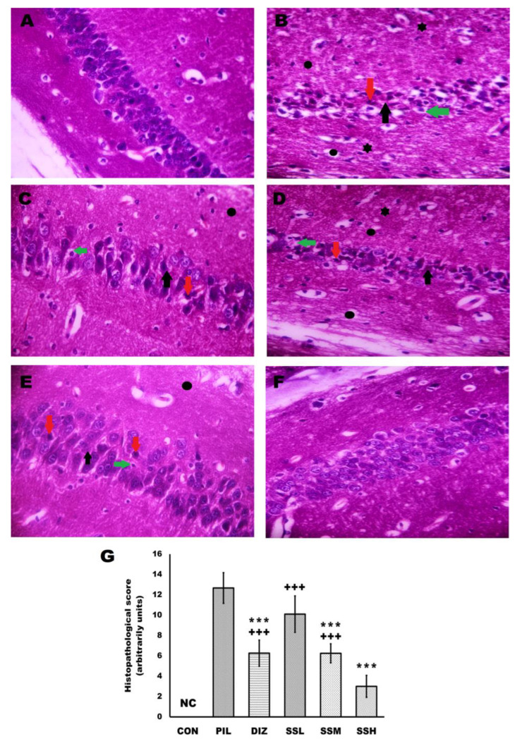
One prevalent neurological disorder is epilepsy. Modulating GABAergic/glutamatergic neurotransmission, Nrf2/HO-1, PI3K/Akt, and TLR-4/NF-B pathways might be a therapeutic strategy for epilepsy. Eight-week-old BALB/c mice were administered 12.5, 25, or 50 mg/kg (-) pseudosemiglabrin orally one hour before inducing epilepsy with an i.p. injection of 360 mg/kg pilocarpine. (-) Pseudosemiglabrin dose-dependently alleviated pilocarpine-induced epilepsy, as revealed by the complete repression of pilocarpine-induced convulsions and 100% survival rate in mice. Furthermore, (-) pseudosemiglabrin significantly enhanced mice's locomotor activities, brain GABA, SLC1A2, GABARα1 levels, glutamate decarboxylase activity, and SLC1A2 and GABARα1mRNA expression while decreasing brain glutamate, SLC6A1, GRIN1 levels, GABA transaminase activity, and SLC6A1 and GRIN1 mRNA expression. These potentials can be due to the suppression of the TLR-4/NF-κB and the enhancement of the Nrf2/HO-1 and PI3K/Akt pathways, as demonstrated by the reduction in TLR-4, NF-κB, IL-1β, TNF-α mRNA expression, MDA, NO, caspase-3, Bax levels, and Bax/Bcl-2 ratio, and the enhancement of Nrf2, HO-1, PI3K, Akt mRNA expression, GSH, Bcl-2 levels, and SOD activity. Additionally, (-) pseudosemiglabrin abrogated the pilocarpine-induced histopathological changes. Interestingly, the (-) pseudosemiglabrin intervention showed a comparable effect to the standard medication, diazepam. Therefore, (-) pseudosemiglabrin can be a promising medication for the management of epilepsy.

Keywords: (-) pseudosemiglabrin; GABAergic and glutamatergic neurotransmission; Nrf2/HO-1 pathway; PI3K/Akt pathway; TLR-4/NF-κB pathway; epilepsy.


Conflict of interest statement

The authors declare no conflict of interest. Moreover, the funders had no role in the design of the study; in the collection, analysis, or interpretation of data; in the writing of the manuscript; or in the decision to publish the results.

Figures

Figure 1
Figure 1
Chemical structure of (-) Pseudosemiglabrin.
Figure 2
Figure 2
(-) Pseudosemiglabrin modulated mice’s brain GABAergic and glutamatergic transmission. The result was expressed as mean ± SD. CON: control group; PIL: pilocarpine-induced convulsion group; DIZ: convulsion-induced group pretreated by single oral dose of diazepam 5 mg/kg; SSL: convulsion-induced group pretreated by single oral dose of (-) pseudosemiglabrin 12.5 mg/kg; SSM: convulsion-induced group pretreated by single oral dose of (-) pseudosemiglabrin 25 mg/kg; SSH: convulsion-induced group pretreated by single oral dose of (-) pseudosemiglabrin 50 mg/kg. * p < 0.05, ** p < 0.01 and *** p < 0.001 (vs. PIL group), + p < 0.05, ++ p < 0.01 and +++ p < 0.001 (vs. SSH group).
Figure 3
Figure 3
(-) Pseudosemiglabrin enhanced the brain GABARα1 and reduced the brain GRIN1 expression in a mouse model of pilocarpine-induced epilepsy. The result was expressed as mean ± SD. CON: control group; PIL: pilocarpine-induced convulsion group; DIZ: convulsion-induced group pretreated by single oral dose of diazepam 5 mg/kg; SSL: convulsion-induced group pretreated by single oral dose of (-) pseudosemiglabrin 12.5 mg/kg; SSM: convulsion-induced group pretreated by single oral dose of (-) pseudosemiglabrin 25 mg/kg; SSH: convulsion-induced group pretreated by single oral dose of (-) pseudosemiglabrin 50 mg/kg. * p < 0.05, ** p < 0.01 and *** p < 0.001 (vs. PIL group), + p < 0.05, ++ p < 0.01 and +++ p < 0.001 (vs. SSH group).
Figure 4
Figure 4
(-) Pseudosemiglabrin enhanced mice’s brain antioxidant system activities. The result was expressed as mean ± SD. CON: control group; PIL: pilocarpine-induced convulsion group; DIZ: convulsion-induced group pretreated by single oral dose of diazepam 5 mg/kg; SSL: convulsion-induced group pretreated by single oral dose of (-) pseudosemiglabrin 12.5 mg/kg; SSM: convulsion-induced group pretreated by single oral dose of (-) pseudosemiglabrin 25 mg/kg; SSH: convulsion-induced group pretreated by single oral dose of (-) pseudosemiglabrin 50 mg/kg. * p < 0.05, ** p < 0.01 and *** p < 0.001 (vs. PIL group), + p < 0.05, ++ p < 0.01 and +++ p < 0.001 (vs. SSH group).
Figure 5
Figure 5
(-) Pseudosemiglabrin-inhibited neuronal apoptosis induced by pilocarpine injection. The result was expressed as mean ± SD. CON: control group; PIL: pilocarpine-induced convulsion group; DIZ: convulsion-induced group pretreated by single oral dose of diazepam 5 mg/kg; SSL: convulsion-induced group pretreated by single oral dose of (-) pseudosemiglabrin 12.5 mg/kg; SSM: convulsion-induced group pretreated by single oral dose of (-) pseudosemiglabrin 25 mg/kg; SSH: convulsion-induced group pretreated by single oral dose of (-) pseudosemiglabrin 50 mg/kg. * p < 0.05, ** p < 0.01 and *** p < 0.001 (vs. PIL group), ++ p < 0.01 and +++ p < 0.001 (vs. SSH group).
Figure 6
Figure 6
(-) Pseudosemiglabrin suppressed brain tissues’ neuro-inflammatory signals induced by pilocarpine injection. The result was expressed as mean ± SD. CON: control group; PIL: pilocarpine-induced convulsion group; DIZ: convulsion-induced group pretreated by single oral dose of diazepam 5 mg/kg; SSL: convulsion-induced group pretreated by single oral dose of (-) pseudosemiglabrin 12.5 mg/kg; SSM: convulsion-induced group pretreated by single oral dose of (-) pseudosemiglabrin 25 mg/kg; SSH: convulsion-induced group pretreated by single oral dose of (-) pseudosemiglabrin 50 mg/kg. * p < 0.05, ** p < 0.01 and *** p < 0.001 (vs. PIL group), + p < 0.05, ++ p < 0.01 and +++ p < 0.001 (vs. SSH group).
Figure 7
Figure 7
(-) Pseudosemiglabrin demotes the brain tissue’s histopathologic changes and histopathological score in pilocarpine-induced convulsion. (A) The control group displayed intact and neatly arranged neurons; (B) the pilocarpine-induced convulsion group displayed severe neuronal degeneration; (C) the convulsion-induced group pretreated by diazepam displayed a mild neuronal degeneration; (DF) convulsion-induced group pretreated by (-) pseudosemiglabrin 12.5, 25, 50 mg/kg, respectively, displayed a dose-dependent mitigation of pilocarpine-induced neurodegeneration to be nearly like the control group in the SSH group. Red arrow: nuclear pyknosis; green arrow: cytoplasmic vacuolation; black arrow: neuronal necrosis; black circle: cerebral congestion; black asterix: inflammatory influx. (G) Histopathological score. NC: no change, *** p < 0.001 (vs. PIL group) and +++ p < 0.001 (vs. SSH group) (H and E, X400).
Figure 8
Figure 8
(-) Pseudosemiglabrin enhanced neuronal survival of the hippocampal CA1 and CA3 cells in pilocarpine-induced convulsion. (I) Nissl’s stain of CA1 hippocampal region; (II) Nissl’s stain of CA3 hippocampal region; (A) control group; (B) pilocarpine-induced convulsion group; (C) convulsion-induced group pretreated by diazepam; (DF) convulsion-induced group pretreated by (-) pseudosemiglabrin 12.5, 25, and 50 mg/kg, respectively; (G) number of surviving hippocampal cells, ** p < 0.01 and *** p < 0.001 (vs. PIL group), ++ p < 0.01 and +++ p < 0.001 (vs. SSH group) (Nissl’s stain, X400).

Similar articles

Cited by

References

    1. Alshabi A.M., Shaikh I.A., Asdaq S.M.B. The antiepileptic potential of Vateria indica Linn in experimental animal models: Effect on brain GABA levels and molecular mechanisms. Saudi J. Biol. Sci. 2022;29:3600–3609. doi: 10.1016/j.sjbs.2022.02.059. - DOI - PMC - PubMed
    1. WHO New WHO Brief Sets out Actions Needed to Improve Lives of People with Epilepsy. [(accessed on 5 January 2023)]; Available online: https://www.who.int/news/item/12-12-2022-new-who-brief-sets-out-actions-....
    1. Simonato M., Löscher W., Cole A.J., Dudek F.E., Engel J., Jr., Kaminski R.M., Loeb J.A., Scharfman H., Staley K.J., Velíšek L. Finding a better drug for epilepsy: Preclinical screening strategies and experimental trial design. Epilepsia. 2012;53:1860–1867. doi: 10.1111/j.1528-1167.2012.03541.x. - DOI - PMC - PubMed
    1. Halassa M.M., Haydon P.G. Integrated brain circuits: Astrocytic networks modulate neuronal activity and behavior. Annu. Rev. Physiol. 2010;72:335. doi: 10.1146/annurev-physiol-021909-135843. - DOI - PMC - PubMed
    1. Bedner P., Dupper A., Hüttmann K., Müller J., Herde M.K., Dublin P., Deshpande T., Schramm J., Häussler U., Haas C.A. Astrocyte uncoupling as a cause of human temporal lobe epilepsy. Brain. 2015;138:1208–1222. doi: 10.1093/brain/awv067. - DOI - PMC - PubMed

MeSH terms