. 2023 Jun 28;24(13):10800.
doi: 10.3390/ijms241310800.

Vps4a Regulates Autophagic Flux to Prevent Hypertrophic Cardiomyopathy

Affiliations

Vps4a Regulates Autophagic Flux to Prevent Hypertrophic Cardiomyopathy

Xiaozhi Huang et al. Int J Mol Sci. .

Abstract

Autophagy has stabilizing functions for cardiomyocytes. Recent studies indicate that an impairment in the autophagy pathway can seriously affect morphology and function, potentially leading to heart failure. However, the role and the underlying mechanism of the endosomal sorting complex required for transport (ESCRT) family protein, in particular the AAA-ATPase vacuolar protein sorting 4a (Vps4a), in regulating myocardial autophagy remains unclear. In the present study, cardiomyocyte-specific Vps4a knockout mice were generated by crossing Vps4aflox/flox (Vps4afl/fl) with Myh6-cre transgenic mice. As a result, we observed a partially dilated left ventricular (LV) chamber, a significant increase in heart weight to body weight ratio (HW/BW), and heart weight to tibial length ratio (HW/TL), hypertrophic cardiomyopathy and early lethality starting at 3 months of age. Hematoxylin-eosin (HE), immunofluorescence assay (IFA), and Western blot (WB) revealed autophagosome accumulation in cardiomyocytes. A transcriptome-based analysis and autophagic flux tracking by AAV-RFP-GFP-LC3 showed that the autophagic flux was blocked in Vps4a knockout cardiomyocytes. In addition, we provided in vitro evidence demonstrating that Vps4a and LC3 were partially co-localized in cardiomyocytes, and the knockdown of Vps4a led to the accumulation of autophagosomes in cardiomyocytes. Similarly, the transfection of cardiomyocytes with adenovirus (Adv) mCherry-GFP-LC3 further indicated that the autophagic flux was blocked in cells with deficient levels of Vps4a. Finally, an electron microscope (EM) showed that the compromised sealing of autophagosome blocked the autophagic flux in Vps4a-depleted cardiomyocytes. These findings revealed that Vps4a contributed to the sealing of autophagosomes in cardiomyocytes. Therefore, we demonstrated that Vps4a deletion could block the autophagic flux, leading to the accumulation of degradation substances and compromised cardiac function. Overall, this study provides insights into a new theoretical basis for which autophagy may represent a therapeutic target for cardiovascular diseases.

Keywords: ESCRT; Vps4a; autophagy; heart; heart failure.


Conflict of interest statement

The authors declare that they have no conflict of interest.

Figures
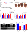
Figure 1
Figure 1
Cardiomyocyte-specific Vps4a deletion affects the anatomy and function of the heart. (A) Kaplan–Meyer curve of Vps4afl/fl Myh6 and Vps4afl/fl mice (n = 60). (B) Bright-field images of the heart from Vps4afl/fl and Vps4afl/fl Myh6 mice. (C,D) Statistical analysis of HW/BW and HW/TL in Vps4afl/fl and Vps4afl/fl Myh6 mice between 3 to 6 months of age (n = 4–8 per group). (EG) Representative transthoracic echocardiography images and statistics of ejection fraction (EF) and left ventricular internal dimension (LVIDd) in Vps4afl/fl and Vps4afl/fl Myh6 mice at 4-months old (n = 5). * p < 0.05, ** p < 0.01, *** p < 0.001, **** p < 0.0001. Significant differences between groups as determined by a two-tailed paired Student’s t-test. Data are expressed as mean and s.e.m. Scale bars: (B) 0.5 mm.
Figure 2
Figure 2
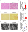
Vps4a-depleted cardiomyocytes showed vacuoles, autophagosomes, and fibrosis. (A) The heart sections of Vps4afl/fl and Vps4afl/fl Myh6 knockout mice were subjected to H&E. Arrows indicate vacuoles in the cardiomyocytes. (B) Statistical analysis number of vacuoles in Vps4afl/fl and Vps4afl/fl Myh6 mice at 3 months of age (n = 3 per group); images in 15 randomly selected visual fields were counted. (C) The heart sections of Vps4afl/fl and Vps4a fl/fl Myh6 knockout mice were subjected to Sirius Red staining. Arrow indicate area of fibrotic cardiomyocytes. (D) Statistical analysis area of fibrosis in Vps4afl/fl and Vps4afl/fl Myh6 mice at 3 months of age (n = 3 per group); images in 15 randomly selected visual fields were counted. (E) The images of autophagosome in Vps4afl/fl Myh6 knockout mice were observed by electron microscope. Arrow indicate autophagosome in the cardiomyocytes. (F) Statistical analysis number of autophagosomes in Vps4afl/fl Myh6 knockout mice (n = 3 per group). Images in 10 randomly selected visual fields were counted. **** p < 0.0001. Significant differences between groups as determined by a two-tailed paired Student’s t-test. Data are expressed as mean and s.e.m. Scale bars: (A,C) 50 μm, (E) 2 μm.
Figure 3
Figure 3
RNA sequencing revealed that autophagy pathway was impaired in Vps4a knockout mice. (A) GO analysis of differentially expressed genes in heart samples of Vps4afl/fl and Vps4afl/fl Myh6 mice, with the top 10 enriched GO terms observed in biological process, cellular component, and molecular function. (B) GO analysis of differentially expressed genes in heart samples from Vps4afl/fl and Vps4afl/fl Myh6 mice, showing GO terms related to autophagy. (C,D) Heat map analysis of differential expression of genes associated with autophagy and mitophagy in the hearts from Vps4afl/fl and Vps4afl/fl Myh6 mice. (Applying the combined criteria of q ≤ 0.05 and fold change ≥1.3, p < 0.05, Bonferroni corrected), (Vps4afl/fl = 3, Vps4afl/fl Myh6 = 6, left: (mild), right: (severe)).
Figure 4
Figure 4
Impaired autophagic flux in Vps4a knockout mice led to abnormal cardiac function. (A,B) Protein expression levels of Vps4a, Myh7, p-AKT, and AKT in heart samples of Vps4afl/fl and Vps4afl/fl Myh6 mice analyzed by Western blot. (C) Protein expression levels of p62 and LC3 in heart samples of Vps4afl/fl and Vps4afl/fl Myh6 mice analyzed by Western blot. (D) Immunofluorescence assay staining of p62, LC3, and α-actinin in heart sections of Vps4afl/fl and Vps4afl/fl Myh6 mice. Arrows indicate staining of p62 and LC3. (E) Autophagic flux tracked by AAV-RFP-GFP-LC3 injected in Vps4afl/fl and Vps4afl/fl Myh6 mice. Arrows indicate autophagosome in the cardiomyocytes. (F) Statistical analysis of autophagosome (yellow) and autolysosome (red) in cardiomyocytes from Vps4afl/fl and Vps4afl/fl Myh6 mice (n = 3 per group) in each experiment, 16 cells were counted. **** p < 0.0001. Significant differences between groups as determined by a two-tailed paired Student’s t-test. Data are expressed as mean and s.e.m. Scale bars: (D, top) 20 μm, (D, bottom, E) 10 μm.
Figure 5
Figure 5
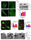
Vps4a is involved in the sealing of autophagosomes within cardiomyocytes and affects the autophagic flux in vitro. (A) Immunofluorescence assay staining of Vps4a and LC3 in H9C2 (top) and primary cardiomyocytes (bottom), with Vps4a and LC3 partially co-localized (arrows). (B) Representative Western blot showing LC3 expression levels in H9C2 cells transfected with Vps4a RNA si and Nc RNA si, with increased expression levels of LC3 II in Vps4a knockdown H9C2 cells. (C), Western blot analysis was performed to measure LC3II/LC3I protein expression. ** p < 0.01 Significant differences between groups as determined by a two-tailed paired Student’s t-test in three independent experiments. (D) Autophagic flux tracked by Adv-mCherry-GFP-LC3 transfected into H9C2 cells. Arrows indicate autophagosome in the cardiomyocytes. (E) Statistic of autophagosome (yellow) and autolysosome (red) in (C), (n = 16) ** p < 0.01. Significant differences between groups as determined by two-way ANOVA with Tukey’s multiple comparisons test. (F) Adv-mCherry-GFP-LC3 was transfected into control and Vps4a knockdown primary cardiomyocytes. Arrows indicate autophagosome in the cardiomyocytes. (G) Statistic of autophagosome (yellow) and autolysosome (red) in (E), (n = 16) **** p < 0.0001. Significant differences between groups as determined by two-way ANOVA with Tukey’s multiple comparisons test. (H) Autophagosome observed by electron microscope in H9C2 cells transfected with Vps4a RNA si and Vps4a RNA sh, showing an incomplete autophagosome atresia. Arrow indicate unclosed autophagosome in the cardiomyocytes. (I) Statistical analysis number of unclosed autophagosome in Vps4a knockdown H9C2 cells. Images in 10 randomly selected visual fields were counted. **** p < 0.0001. Significant differences between groups as determined by a two-tailed paired Student’s t-test. Data are expressed as mean and s.e.m. Scale bars: (A,D,F) 10 μm, (H) 1 μm.
Figure 6
Figure 6
Schematic showing that Vps4a controls autophagic flux to regulate cardiac function. During the process of autophagosome formation, Vps4a is involved in the sealing of the autophagosome. When Vps4a is conditionally deleted in a mouse model, the autophagic flux in cardiomyocytes is blocked, which impairs the degradation of intracellular components and leads to cardiomyocyte death and fibrosis. The reduced cardiac function and heart failure results in early lethality in Vps4a knockout mice.

Similar articles

Cited by

References

    1. Klionsky D.J., Emr S.D. Autophagy as a Regulated Pathway of Cellular Degradation. Science. 2000;290:1717–1721. doi: 10.1126/science.290.5497.1717. - DOI - PMC - PubMed
    1. Galluzzi L., Baehrecke E.H., Ballabio A., Boya P., Bravo San Pedro J.M., Cecconi F., Choi A.M., Chu C.T., Codogno P., Colombo M.I., et al. Molecular definitions of autophagy and related processes. EMBO J. 2017;36:1811–1836. doi: 10.15252/embj.201796697. - DOI - PMC - PubMed
    1. Levine B., Klionsky D.J. Development by Self-Digestion: Molecular Mechanisms and Biological Functions of Autophagy. Dev Cell. 2004;6:463–477. doi: 10.1016/S1534-5807(04)00099-1. - DOI - PubMed
    1. Ichimiya T., Yamakawa T., Hirano T., Yokoyama Y., Hayashi Y., Hirayama D., Wagatsuma K., Itoi T., Nakase H. Autophagy and Autophagy-Related Diseases: A Review. Int. J. Mol. Sci. 2020;21:8974. doi: 10.3390/ijms21238974. - DOI - PMC - PubMed
    1. Bravo-San Pedro J.M., Kroemer G., Galluzzi L. Autophagy and Mitophagy in Cardiovascular Disease. Circ. Res. 2017;120:1812–1824. doi: 10.1161/CIRCRESAHA.117.311082. - DOI - PubMed

LinkOut - more resources