Randomized Controlled Trial
. 2023 Jun 28;24(13):10815.
doi: 10.3390/ijms241310815.

The Oral Transglutaminase 2 Inhibitor ZED1227 Accumulates in the Villous Enterocytes in Celiac Disease Patients during Gluten Challenge and Drug Treatment

Affiliations
Randomized Controlled Trial

The Oral Transglutaminase 2 Inhibitor ZED1227 Accumulates in the Villous Enterocytes in Celiac Disease Patients during Gluten Challenge and Drug Treatment

Jorma Isola et al. Int J Mol Sci. .

Abstract

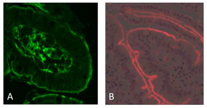
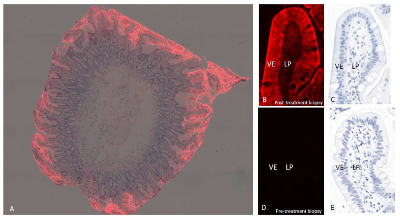
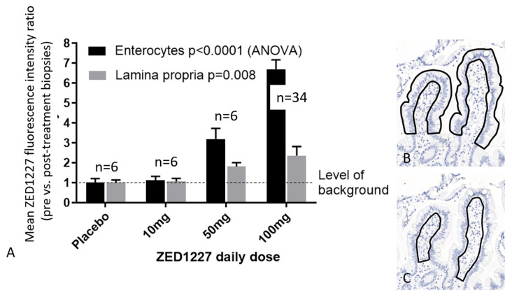
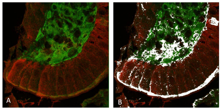
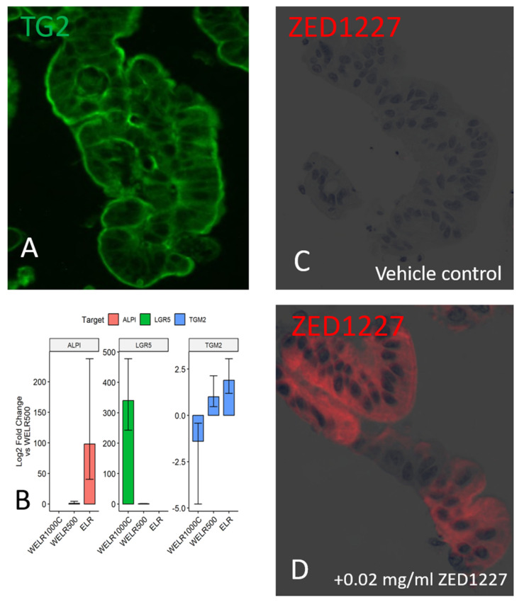
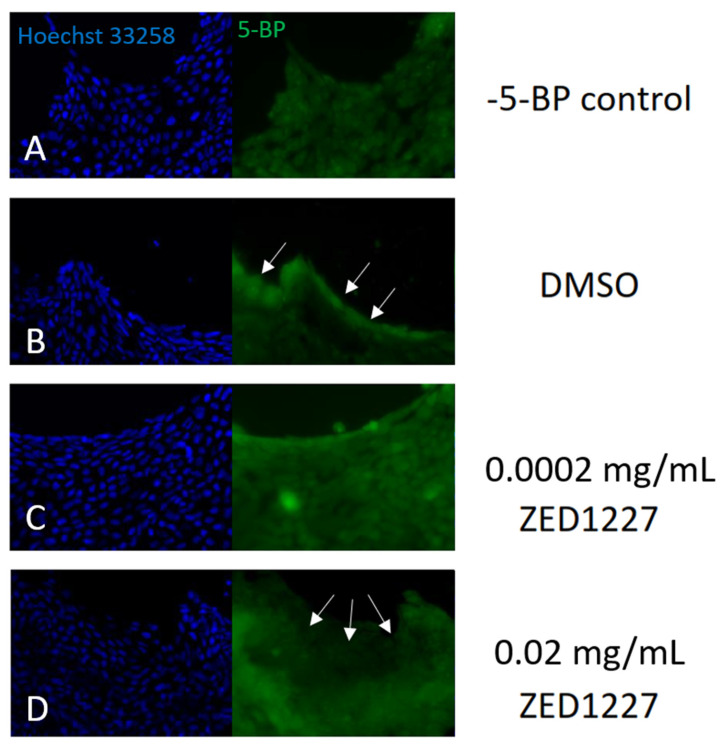
The enzyme transglutaminase 2 (TG2) plays a key role in celiac disease (CeD) pathogenesis. Active TG2 is located mainly extracellularly in the lamina propria but also in the villous enterocytes of the duodenum. The TG2 inhibitor ZED1227 is a promising drug candidate for treating CeD and is designed to block the TG2-catalyzed deamidation and crosslinking of gliadin peptides. Our aim was to study the accumulation of ZED1227 after oral administration of the drug. We studied duodenal biopsies derived from a phase 2a clinical drug trial using an antibody that detects ZED1227 when bound to the catalytic center of TG2. Human epithelial organoids were studied in vitro for the effect of ZED1227 on the activity of TG2 using the 5-biotin-pentylamine assay. The ZED1227-TG2 complex was found mainly in the villous enterocytes in post-treatment biopsies. The signal of ZED1227-TG2 was strongest in the luminal epithelial brush border, while the intensity of the signal in the lamina propria was only ~20% of that in the villous enterocytes. No signal specific to ZED1227 could be detected in pretreatment biopsies or in biopsies from patients randomized to the placebo treatment arm. ZED1227-TG2 staining co-localized with total TG2 and native and deamidated gliadin peptides on the enterocyte luminal surface. Inhibition of TG2 activity by ZED1227 was demonstrated in epithelial organoids. Our findings suggest that active TG2 is present at the luminal side of the villous epithelium and that inhibition of TG2 activity by ZED1227 occurs already there before gliadin peptides enter the lamina propria.

Keywords: brush border; celiac disease therapy; enterocyte; gliadin; gluten.


Conflict of interest statement

J.I. is the owner of Jilab Inc. M.H. and R.P. are employees and shareholders of Zedira and co-inventors of a patent covering the ZED1227 compound. T.Z., R.M. and R.G. are employees of DrFalkPharma Gmbh. M.M. is the owner and Chair of Board at Maki HealthTech Ltd. (MHT). MHT receives management/Advisory Affiliation fees from Dr. Falk Pharma, Topas Therapeutics, Calypso Biotech, Vaccitech, ImmunogenX, and Equillium; holds patents licensed to Labsystems Diagnostics, from which MHT receives royalties via Tampere University Hospital.

Figures

Figure 1
Figure 1
Immunofluorescence staining of transglutaminase 2 (using mab CUB 7402, green fluorescence) and gliadin peptides (mab A161, red fluorescence) in a duodenal biopsy from a CeD patient. The majority of the total TG2 is localized near the basement membrane and in the lamina propria. The luminal surface of the villous epithelium is labeled with equally strong intensity (panel (A)). Gliadin peptides (33-mer, non-deamidated, and deamidated) also localize on the luminal surface of the villous epithelium (panel (B)). Original magnification: ×200.
Figure 2
Figure 2
Localization of ZED1227 by mab A083 immunofluorescence (red) in a duodenal biopsy from a ZED1227-treated patient at low magnification (panel (A), image blended with hematoxylin counterstain). The signal accumulation in the villous epithelium is evident. At higher magnification, a post-treatment (100 mg/day) biopsy shows the labeling localized mainly in the apical surface of the villous epithelium (VE) (panel (B), counterstain in panel (C)). Lower-intensity ZED1227-TG2 labeling can be seen in the lamina propria (LP). The fluorescence signal is absent in a pre-treatment biopsy from the same patient (panels (D,E)).
Figure 3
Figure 3
Comparison of quantified ZED1227-TG2 fluorescence intensities in the villous epithelium and lamina propria in the patient groups who received placebo, 10 mg, 50 mg, or 100 mg of ZED1227. The last oral drug dose was given 24 h before endoscopy and biopsy sampling (panel (A)). Panels (B,C) illustrate the regions drawn to define the epithelial and lamina propria measurement areas. Shown are the average fluorescence intensity ratios (pre- vs. post-treatment biopsies of each patient stained on the same slide). Original magnification in panels B and C, ×100.
Figure 4
Figure 4
Confocal microscopy imaging of total TG2 (mab CUB 7402, in green) and ZED1227 (mab A083, in red) by double immunofluorescence staining (panel (A)). White pixels in panel (B) show the areas with statistically significant co-localization defined by the ImageJ/FIJI image analysis co-localization algorithm. Original magnification: ×600.
Figure 5
Figure 5
Panel (A) shows the immunofluorescence localization of total TG2 in a lamina-propria- free intestinal human epithelial organoid. Panel (B) shows the expression level of TG2 mRNA, as determined by RT–qPCR. The mRNA level is increased along with villous differentiation (using the ELR medium). The increase parallels with that of the mature enterocyte marker (ALPI), while the mRNA level of the stem cell marker LGR5 is decreased. Plots with whiskers represent the mean and standard error (n = 2). Panels (C,D) demonstrate the localization of ZED1227 in the epithelial organoid cells before (panel (C)) and after adding 0.02 mg/mL of ZED1227 in the culture medium (red fluorescence).
Figure 6
Figure 6
TG2 activity is demonstrated with 5-biotin pentylamine labeling in a cultured human epithelial organoid. The TG2 inhibitor ZED1227 (0.02 mg/mL) caused a reduction of 5-BP staining intensity in the apical epithelial surface compared to the DMSO control (arrows). Panel (A) (negative control); panel (B) (DMSO control; no TG2 inhibitor added); panels (C,D) (with ZED1227 0.002 mg/mL and 0.02 mg/mL added, respectively).

References

    1. Kivelä L., Caminero A., Leffler D.A., Pinto-Sanchez M.I., Tye-Din J.A., Lindfors K. Current and emerging therapies for coeliac disease. Nat. Rev. Gastroenterol. Hepatol. 2021;18:181–195. doi: 10.1038/s41575-020-00378-1. - DOI - PubMed
    1. Korponay-Szabó I.R., Halttunen T., Szalai Z., Laurila K., Király R., Kovács J.B., Fésüs L., Mäki M. In vivo targeting of intestinal and extraintestinal transglutaminase 2 by coeliac autoantibodies. Gut. 2004;53:641–648. doi: 10.1136/gut.2003.024836. - DOI - PMC - PubMed
    1. Gatti S., Rossi M., Alfonsi S., Mandolesi A., Cobellis G., Catassi C. Beyond the Intestinal Celiac Mucosa: Diagnostic Role of Anti-TG2 Deposits, a Systematic Review. Front. Med. 2014;1:9. doi: 10.3389/fmed.2014.00009. - DOI - PMC - PubMed
    1. Iversen R., Amundsen S.F., Kleppa L., du Pré M.F., Stamnaes J., Sollid L.M. Evidence That Pathogenic Transglutaminase 2 in Celiac Disease Derives from Enterocytes. Gastroenterology. 2020;159:788–790. doi: 10.1053/j.gastro.2020.04.018. - DOI - PubMed
    1. Elmentaite R., Kumasaka N., Roberts K., Fleming A., Dann E., King H.W., Kleshchevnikov V., Dabrowska M., Pritchard S., Bolt L., et al. Cells of the human intestinal tract mapped across space and time. Nature. 2021;597:250–255. doi: 10.1038/s41586-021-03852-1. - DOI - PMC - PubMed

Publication types