Review
. 2023 Jun 28;24(13):10825.
doi: 10.3390/ijms241310825.

Building a Scaffold for Arteriovenous Fistula Maturation: Unravelling the Role of the Extracellular Matrix

Affiliations
Review

Building a Scaffold for Arteriovenous Fistula Maturation: Unravelling the Role of the Extracellular Matrix

Suzanne L Laboyrie et al. Int J Mol Sci. .

Abstract

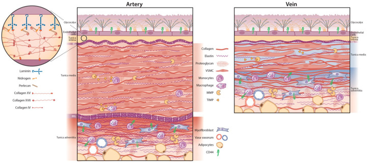
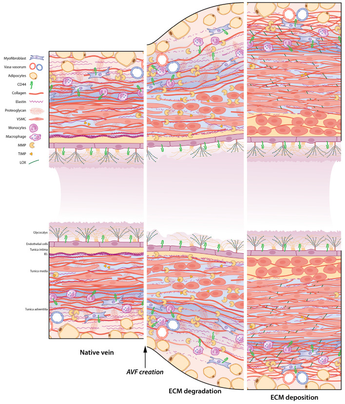
Vascular access is the lifeline for patients receiving haemodialysis as kidney replacement therapy. As a surgically created arteriovenous fistula (AVF) provides a high-flow conduit suitable for cannulation, it remains the vascular access of choice. In order to use an AVF successfully, the luminal diameter and the vessel wall of the venous outflow tract have to increase. This process is referred to as AVF maturation. AVF non-maturation is an important limitation of AVFs that contributes to their poor primary patency rates. To date, there is no clear overview of the overall role of the extracellular matrix (ECM) in AVF maturation. The ECM is essential for vascular functioning, as it provides structural and mechanical strength and communicates with vascular cells to regulate their differentiation and proliferation. Thus, the ECM is involved in multiple processes that regulate AVF maturation, and it is essential to study its anatomy and vascular response to AVF surgery to define therapeutic targets to improve AVF maturation. In this review, we discuss the composition of both the arterial and venous ECM and its incorporation in the three vessel layers: the tunica intima, media, and adventitia. Furthermore, we examine the effect of chronic kidney failure on the vasculature, the timing of ECM remodelling post-AVF surgery, and current ECM interventions to improve AVF maturation. Lastly, the suitability of ECM interventions as a therapeutic target for AVF maturation will be discussed.

Keywords: AVF maturation; arteriovenous fistula; extracellular matrix; vascular remodelling.


Conflict of interest statement

The authors declare no conflict of interest.

Figures

Figure 1
Figure 1
Overview of the arterial versus venous vessel layers and their respective ECM. The luminal side of both vessel types contains an intimal layer with endothelial cells (ECs) covered by the glycocalyx. The basal lamina is a mesh-like structure situated underneath the ECs. The internal elastic lamina (IEL) separates the tunica intima and media. The arterial (left panel) tunica media is thicker and more elastic than its venous counterpart (right panel) with more vascular smooth muscle cells (VSMCs), organised with elastin into a contractile-elastic unit. Arteries have a prominent external elastic lamina (EEL). Veins are less muscular with a lower elastin-to-collagen ratio. The tunica adventitia contains collagen-producing (myo) fibroblasts, surrounded by perivascular adipose tissue and the vasa vasorum: a capillary network of minor blood vessels. Matrix metalloproteinases (MMPs) are proteinases that regulate ECM degradation and extend into the media and adventitia. They are inhibited by tissue inhibitors of metalloproteinases (TIMPs). CD44 is a cell-surface glycoprotein receptor found in the venous and arterial adventitial layer and venous tunica intima. CD44 is expressed by ECs and can bind ECM components, such as collagen, fibronectin, MMPs, and hyaluronic acid. ECM = extracellular matrix.
Figure 2
Figure 2
ECM remodelling after AVF creation. The native vein (left panel) requires rebuilding its vascular scaffold to facilitate AVF maturation. The venous outflow tract undergoes ECM degradation (middle panel) and rebuilding of the ECM framework through ECM deposition (right panel). After AVF creation, the intimal layer and glycocalyx are damaged, and increased wall shear stress induces outward remodelling. Degradation of the ECM is facilitated by increased MMP (matrix metalloprotease) production, degrading collagen and elastin. Proliferating VSMCs ensure outward remodelling and wall thickening. Eventually, VSMCs migrate into the intima, where they proliferate and form intimal hyperplasia. During ECM deposition, collagen is produced by VSMCs and myofibroblasts. LOX (lysyl oxidase) cross-links elastin and collagen into their respective fibre formations.
Figure 3
Figure 3
Differential ECM deposition in human AVFs. Samples were stained with Masson Trichrome. ECM is shown in blue, and smooth muscle cells are stained red. Scale bars are 100 µm in the bottom images and 500 µm in the inlay. (A) shows an AVF with little outward remodelling and a lot of ECM deposition in the IH, as shown in the inlay, indicative of a fibrotic AVF. (B) AVF with wall thickening and some ECM deposition amongst well-organised muscle fibres. (C) AVF with excessive OR and little ECM deposition, indicative of aneurysm formation. AVF = arteriovenous fistula, ECM = extracellular matrix, IH = intimal hyperplasia, OR = outward remodelling.

Similar articles

Cited by

References

    1. Brescia M.J., Cimino J.E., Appel K., Hurwich B.J. Chronic hemodialysis using venipuncture and a surgically created arteriovenous fistula. N. Engl. J. Med. 1966;275:1089–1092. doi: 10.1056/NEJM196611172752002. - DOI - PubMed
    1. Schmidli J., Widmer M.K., Basile C., de Donato G., Gallieni M., Gibbons C.P., Haage P., Hamilton G., Hedin U., Kamper L., et al. Editor’s Choice—Vascular Access: 2018 Clinical Practice Guidelines of the European Society for Vascular Surgery (ESVS) Eur. J. Vasc. Endovasc. Surg. 2018;55:757–818. doi: 10.1016/j.ejvs.2018.02.001. - DOI - PubMed
    1. Lok C.E., Huber T.S., Lee T., Shenoy S., Yevzlin A.S., Abreo K., Allon M., Asif A., Astor B.C., Glickman M.H., et al. KDOQI Clinical Practice Guideline for Vascular Access: 2019 Update. Am. J. Kidney Dis. 2020;75:S1–S164. doi: 10.1053/j.ajkd.2019.12.001. - DOI - PubMed
    1. Astor B.C., Eustace J.A., Powe N.R., Klag M.J., Fink N.E., Coresh J., Josef Coresh for the CHOICE Study Type of Vascular Access and Survival among Incident Hemodialysis Patients: The Choices for Healthy Outcomes in Caring for ESRD (CHOICE) Study. J. Am. Soc. Nephrol. 2005;16:1449–1455. doi: 10.1681/ASN.2004090748. - DOI - PubMed
    1. Banerjee T., Kim S.J., Astor B., Shafi T., Coresh J., Powe N.R. Vascular Access Type, Inflammatory Markers, and Mortality in Incident Hemodialysis Patients: The Choices for Healthy Outcomes in Caring for End-Stage Renal Disease (CHOICE) Study. Am. J. Kidney Dis. 2014;64:954–961. doi: 10.1053/j.ajkd.2014.07.010. - DOI - PMC - PubMed