. 2023 Jun 29;24(13):10877.
doi: 10.3390/ijms241310877.

Bypass of Abasic Site-Peptide Cross-Links by Human Repair and Translesion DNA Polymerases

Affiliations

Bypass of Abasic Site-Peptide Cross-Links by Human Repair and Translesion DNA Polymerases

Anna V Yudkina et al. Int J Mol Sci. .

Abstract

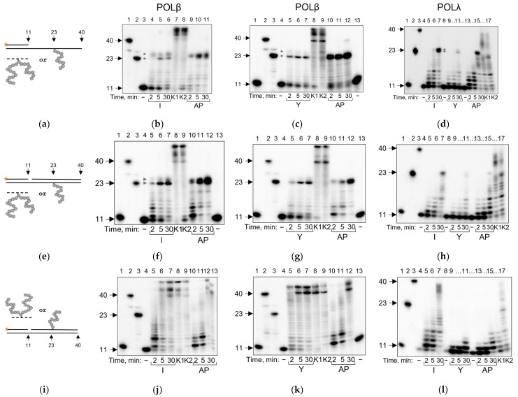
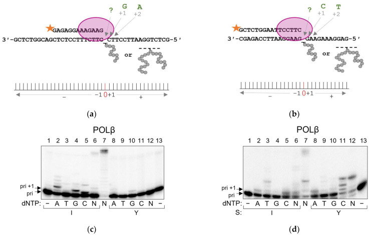
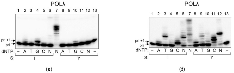
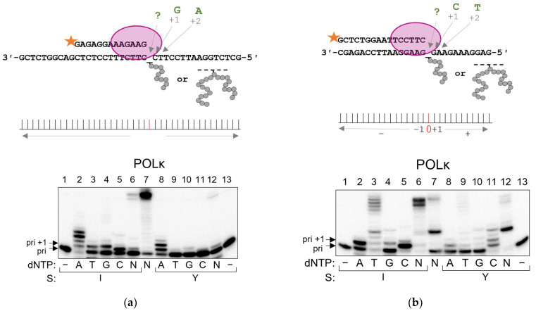
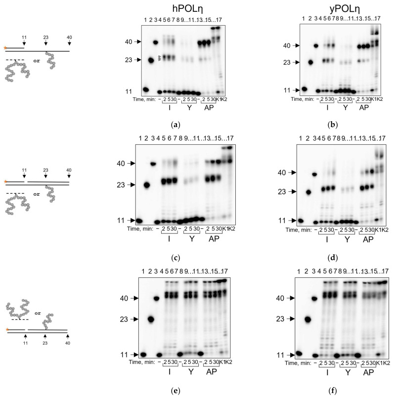
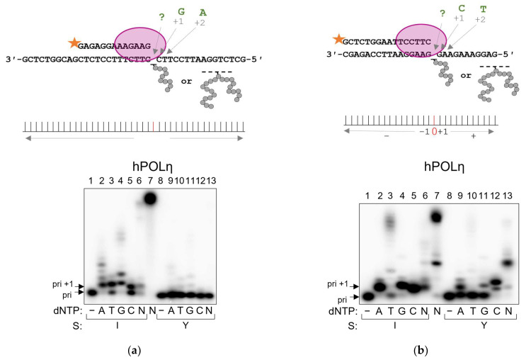
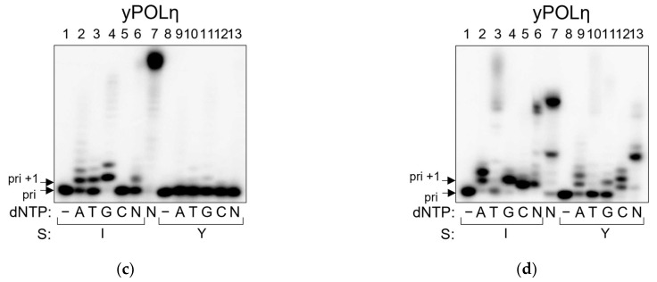
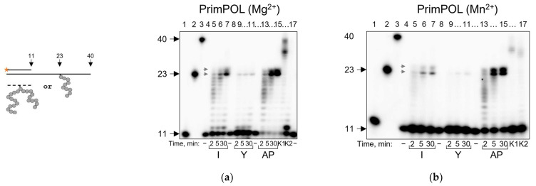
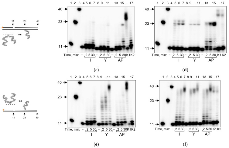
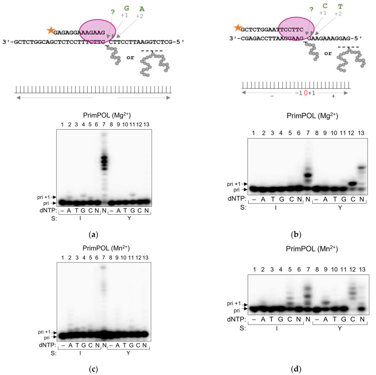
DNA-protein cross-links remain the least-studied type of DNA damage. Recently, their repair was shown to involve proteolysis; however, the fate of the peptide remnant attached to DNA is unclear. Particularly, peptide cross-links could interfere with DNA polymerases. Apurinuic/apyrimidinic (AP) sites, abundant and spontaneously arising DNA lesions, readily form cross-links with proteins. Their degradation products (AP site-peptide cross-links, APPXLs) are non-instructive and should be even more problematic for polymerases. Here, we address the ability of human DNA polymerases involved in DNA repair and translesion synthesis (POLβ, POLλ, POLη, POLκ and PrimPOL) to carry out synthesis on templates containing AP sites cross-linked to the N-terminus of a 10-mer peptide (APPXL-I) or to an internal lysine of a 23-mer peptide (APPXL-Y). Generally, APPXLs strongly blocked processive DNA synthesis. The blocking properties of APPXL-I were comparable with those of an AP site, while APPXL-Y constituted a much stronger obstruction. POLη and POLκ demonstrated the highest bypass ability. DNA polymerases mostly used dNTP-stabilized template misalignment to incorporate nucleotides when encountering an APPXL. We conclude that APPXLs are likely highly cytotoxic and mutagenic intermediates of AP site-protein cross-link repair and must be quickly eliminated before replication.

Keywords: AP sites; DNA damage; DNA lesion bypass; DNA polymerases; DNA repair; DNA–peptide cross-links; mutagenesis; translesion synthesis.


Conflict of interest statement

The authors declare no conflict of interest.

Figures

Figure 1
Figure 1
Scheme of APPXL substrates. E. coli Fpg and human OGG1 are cross-linked by NaBH4 to a DNA duplex containing a single 8-oxoguanine residue; as a result, a stable AP site–protein cross-link is formed. After extensive trypsinolysis, Fpg yields an N-terminally cross-linked 10-mer peptide (APPXL-I) and OGG1, an internally cross-linked 23-mer peptide (APPXL-Y). Structures shown are those of the NaBH4-trapped AP site–protein cross-links with E. coli Fpg [40] and human OGG1 [41]. In the peptide sequences, a tilde marks the cross-linking site.
Figure 2
Figure 2
Running start synthesis by POLβ and POLλ on APPXL-containing substrates. Primer–template substrate, and adduct in the template (a); representative gels showing extension for POLβ and APPXL-I (b); POLβ and APPXL-Y (c); POLλ and both APPXLs (d). Primer–template–downstream strand substrate, and adduct in the template (e); representative gels showing extension for POLβ and APPXL-I (f); POLβ and APPXL-Y (g) and POLλ and both APPXLs (h). Primer–template–downstream strand substrate, and adduct in the downstream strand (i); representative gels showing extension for POLβ and APPXL-I (j); POLβ and APPXL-Y (k) and POLλ and both APPXLs (l). The nature of the substrate and reaction time are indicated below the gel image; I, APPXL-I; Y, APPXL-Y; AP, natural AP site. K1, undamaged primer–template substrate; K2, undamaged primer–downstream strand–template substrate; “−”, no enzyme added. In all gels: lanes 1–3, size markers with lengths corresponding to the primer (11 nt), full-size product (40 nt) and primer extended to the site of cross-linking (23 nt). Gray arrows indicate synthesis pause points.
Figure 3
Figure 3
Incorporation preference of POLβ and POLλ in standing start synthesis on APPXL-containing substrates. Schemes of the substrates are shown above the gel images. Pyrimidine-rich template (a); representative gels for nucleotide incorporation by POLβ (c) and POLλ (e). Purine-rich template (b); representative gels for nucleotide incorporation by POLβ (d) and POLλ (f). The nature of the substrate and dNTP are indicated below the gel image; I, APPXL-I; Y, APPXL-Y; N, mixture of all four dNTPs. K1, undamaged primer–template substrate; K2, undamaged primer–downstream strand–template substrate. In all gels: lane 7, undamaged substrate; “−”, no enzyme added.
Figure 3
Figure 3
Incorporation preference of POLβ and POLλ in standing start synthesis on APPXL-containing substrates. Schemes of the substrates are shown above the gel images. Pyrimidine-rich template (a); representative gels for nucleotide incorporation by POLβ (c) and POLλ (e). Purine-rich template (b); representative gels for nucleotide incorporation by POLβ (d) and POLλ (f). The nature of the substrate and dNTP are indicated below the gel image; I, APPXL-I; Y, APPXL-Y; N, mixture of all four dNTPs. K1, undamaged primer–template substrate; K2, undamaged primer–downstream strand–template substrate. In all gels: lane 7, undamaged substrate; “−”, no enzyme added.
Figure 4
Figure 4
Running start synthesis by POLκ on APPXL-containing substrates. Schemes of the substrates are shown to the left of the gel images. Primer–template substrate, and adduct in the template: (a) APPXL-I; (b) APPXL-Y. Primer–template–downstream strand substrate, and adduct in the template: (c) APPXL-I; (d) APPXL-Y. Primer–template–downstream strand substrate, and adduct in the downstream strand: (e) APPXL-I; (f) APPXL-Y. The nature of the substrate and reaction time are indicated below the gel image; I, APPXL-I; Y, APPXL-Y; AP, natural AP site. K1, undamaged primer–template substrate; K2, undamaged primer–downstream strand–template substrate; “−”, no enzyme added. In all gels: lanes 1–3, size markers with lengths corresponding to the primer (11 nt), full-size product (40 nt) and primer extended to the site of cross-linking (23 nt). Gray arrows indicate synthesis pause points.
Figure 5
Figure 5
Incorporation preference of POLκ in standing start synthesis on APPXL-containing substrates. Schemes of the substrates are shown above the gel images. (a) Pyrimidine-rich template, (b) purine-rich template. The nature of the substrate and dNTP are indicated below the gel image; I, APPXL-I; Y, APPXL-Y; N, mixture of all four dNTPs. K1, undamaged primer–template substrate; K2, undamaged primer–downstream strand–template substrate. In all gels: lane 7, undamaged substrate; “−“, no enzyme added.
Figure 6
Figure 6
Running start synthesis by hPOLη and yPOLη on APPXL-containing substrates. Schemes of the substrates are shown to the left of the gel images. Primer–template substrate, and adduct in the template: (a) hPOLη; (b) yPOLη. Primer–template–downstream strand substrate, and adduct in the template: (c) hPOLη; (d) yPOLη. Primer–template–downstream strand substrate, and adduct in the downstream strand: (e) hPOLη; (f) yPOLη. The nature of the substrate and reaction time are indicated below the gel image; I, APPXL-I; Y, APPXL-Y; AP, natural AP site. K1, undamaged primer–template substrate; K2, undamaged primer–downstream strand–template substrate; “−”, no enzyme added. In all gels: lanes 1–3, size markers with lengths corresponding to the primer (11 nt), full-size product (40 nt) and primer extended to the site of cross-linking (23 nt). Gray arrows indicate synthesis pause points.
Figure 7
Figure 7
Incorporation preference of hPOLη and yPOLη in standing start synthesis on APPXL-containing substrates. Schemes of the substrates are shown above the gel images. Pyrimidine-rich template: (a) hPOLη; (c) yPOLη. Purine-rich template: (b) hPOLη; (d) yPOLη. The nature of the substrate and dNTP are indicated below the gel image; I, APPXL-I; Y, APPXL-Y; N, mixture of all four dNTPs. K1, undamaged primer–template substrate; K2, undamaged primer–downstream strand–template substrate. In all gels: lane 7, undamaged substrate; “−“, no enzyme added.
Figure 7
Figure 7
Incorporation preference of hPOLη and yPOLη in standing start synthesis on APPXL-containing substrates. Schemes of the substrates are shown above the gel images. Pyrimidine-rich template: (a) hPOLη; (c) yPOLη. Purine-rich template: (b) hPOLη; (d) yPOLη. The nature of the substrate and dNTP are indicated below the gel image; I, APPXL-I; Y, APPXL-Y; N, mixture of all four dNTPs. K1, undamaged primer–template substrate; K2, undamaged primer–downstream strand–template substrate. In all gels: lane 7, undamaged substrate; “−“, no enzyme added.
Figure 8
Figure 8
Running start synthesis by PrimPOL on APPXL-containing substrates. Schemes of the substrates are shown to the left of the gel images. Primer–template substrate, and adduct in the template: APPXL-I and APPXL-Y in the presence of Mg2+ (a) or Mn2+ (b). Primer–template–downstream strand substrate, and adduct in the template: APPXL-I and APPXL-Y in the presence of Mg2+ (c) or Mn2+ (d). Primer–template–downstream strand substrate, and adduct in the downstream strand: APPXL-I and APPXL-Y in the presence of Mg2+ (e) or Mn2+ (f). The nature of the substrate and reaction time are indicated below the gel image; I, APPXL-I; Y, APPXL-Y; AP, natural AP site. K1, undamaged primer–template substrate; K2, undamaged primer–downstream strand–template substrate; “−”, no enzyme added. In all gels: lanes 1–3, size markers with lengths corresponding to the primer (11 nt), full-size product (40 nt) and primer extended to the site of cross-linking (23 nt). Gray arrows indicate synthesis pause points.
Figure 8
Figure 8
Running start synthesis by PrimPOL on APPXL-containing substrates. Schemes of the substrates are shown to the left of the gel images. Primer–template substrate, and adduct in the template: APPXL-I and APPXL-Y in the presence of Mg2+ (a) or Mn2+ (b). Primer–template–downstream strand substrate, and adduct in the template: APPXL-I and APPXL-Y in the presence of Mg2+ (c) or Mn2+ (d). Primer–template–downstream strand substrate, and adduct in the downstream strand: APPXL-I and APPXL-Y in the presence of Mg2+ (e) or Mn2+ (f). The nature of the substrate and reaction time are indicated below the gel image; I, APPXL-I; Y, APPXL-Y; AP, natural AP site. K1, undamaged primer–template substrate; K2, undamaged primer–downstream strand–template substrate; “−”, no enzyme added. In all gels: lanes 1–3, size markers with lengths corresponding to the primer (11 nt), full-size product (40 nt) and primer extended to the site of cross-linking (23 nt). Gray arrows indicate synthesis pause points.
Figure 9
Figure 9
Incorporation preference of PrimPOL in standing start synthesis on APPXL-containing substrates. Schemes of the substrates are shown above the gel images. Pyrimidine-rich template (a) in the presence of Mg2+ and (c) in the presence of Mn2+. Purine-rich template (b) in the presence of Mg2+ and (d) in the presence of Mn2+. The nature of the substrate and dNTP are indicated below the gel image; I, APPXL-I; Y, APPXL-Y; N, mixture of all four dNTPs. K1, undamaged primer–template substrate; K2, undamaged primer–downstream strand–template substrate. In all gels: lane 7, undamaged substrate; “−“, no enzyme added.

Similar articles

Cited by

References

    1. Lindahl T., Nyberg B. Rate of depurination of native deoxyribonucleic acid. Biochemistry. 1972;11:3610–3618. doi: 10.1021/bi00769a018. - DOI - PubMed
    1. Atamna H., Cheung I., Ames B.N. A method for detecting abasic sites in living cells: Age-dependent changes in base excision repair. Proc. Natl. Acad. Sci. USA. 2000;97:686–691. doi: 10.1073/pnas.97.2.686. - DOI - PMC - PubMed
    1. Guan L., Greenberg M.M. DNA interstrand cross-link formation by the 1,4-dioxobutane abasic lesion. J. Am. Chem. Soc. 2009;131:15225–15231. doi: 10.1021/ja9061695. - DOI - PMC - PubMed
    1. Ghosh S., Greenberg M.M. Correlation of thermal stability and structural distortion of DNA interstrand cross-links produced from oxidized abasic sites with their selective formation and repair. Biochemistry. 2015;54:6274–6283. doi: 10.1021/acs.biochem.5b00860. - DOI - PMC - PubMed
    1. Greenberg M.M., Weledji Y.N., Kroeger K.M., Kim J. In vitro replication and repair of DNA containing a C2′-oxidized abasic site. Biochemistry. 2004;43:15217–15222. doi: 10.1021/bi048360c. - DOI - PubMed

LinkOut - more resources