Review
. 2023 Jun 29;24(13):10882.
doi: 10.3390/ijms241310882.

Microfluidics and Organoids, the Power Couple of Developmental Biology and Oncology Studies

Affiliations
Review

Microfluidics and Organoids, the Power Couple of Developmental Biology and Oncology Studies

Laura Ann Hetzel et al. Int J Mol Sci. .

Abstract

Organoids are an advanced cell model that hold the key to unlocking a deeper understanding of in vivo cellular processes. This model can be used in understanding organ development, disease progression, and treatment efficacy. As the scientific world embraces the model, it must also establish the best practices for cultivating organoids and utilizing them to the greatest potential in assays. Microfluidic devices are emerging as a solution to overcome the challenges of organoids and adapt assays. Unfortunately, the various applications of organoids often depend on specific features in a device. In this review, we discuss the options and considerations for features and materials depending on the application and development of the organoid.

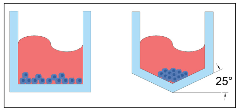
Keywords: microfluidic device; organoid filtering; organoid formation; organoids.


Conflict of interest statement

The authors declare no conflict of interest.

Figures

Figure 1
Figure 1
A typical three channel device, with three inlets and three outlets. The colors help identify individual channels. Cells are typically in the center channel, allowing media, drugs, and stains to be perfused through the outer two channels. The left image is a simulated model of the device and on the right is a drawing of a comparable three channel device.
Figure 2
Figure 2
Left: A three channel device with hexagonal posts instead of solid walls to enclose each channel. Right: A close up showing the shape of the hexagonal posts supporting a hydrogel in the center channel.
Figure 3
Figure 3
An organoid will be either trapped or released from a trapping blockade depending on the direction of the flow of media, as indicated with the arrows. In the bottom scenario, the organoid is trapped in a rectangular blockade when the media is flowing from the direction of the large opening of the blockade toward the smaller opening. When the flow is reversed, as shown in the top scenario, the organoid is released from the blockade.
Figure 4
Figure 4
Microfluidic device with five wells: the deeper ones to serve as media reservoirs and the shallower ones to house the organoids. The channels connecting the wells are layered to provide enough flow for the exchange of nutrients and waste while limiting the shear stresses for optimal growth of the brain organoids.
Figure 5
Figure 5
Regardless of the full device layout, the bottom should provide a shallow well for media, with a mesh above it so that the media can get into the larger organoid wells that are on the top. The mesh prevents the organoid from sitting on the bottom of a well and allows for volume control. On the right, organoids represented by yellow spheres are shown resting on the mesh in the well.
Figure 6
Figure 6
The U-shaped traps on the top row will trap organoids of any size (left two U traps), as well as single cells (right U trap). The open traps on the bottom row allow the smaller organoids and single cells to pass as indicated with the green check mark arrows and prohibiting the larger organoids to pass as indicated with the red forbidden arrow, acting as a filter for organoids of a specified size.
Figure 7
Figure 7
Microfluidic device designed to trap the organoid with media flow (indicated with solid red arrow), provide a hydrogel matrix (striped, orange) and expose the organoid to two different sources of media (cross hatched green and blue) to expose opposing sides of the organoid to different chemical gradients.
Figure 8
Figure 8
Basic manufacturing process of Poly(dimethylsiloxane) (PDMS) devices. (A) design the device in computer-aided drafting (CAD) software and translate to a negative mold in the software. (B) Use photoresist layer (red hatch) on a silicon wafer (solid green) strategically blocked from light with a transparency film (solid gray) (C) After light treatment, the reusable negative mold silicon wafer is ready for use. (D) Curing a 10:1 ratio of base and curing agent in a vacuum forms the device shape (blue double line hatch). (E) The device is cut out of the mold and holes punctured (F) PDMS is reversibly attached to a glass slide (solid dark gray) with heat. Figure partially created with BioRender.com.
Figure 9
Figure 9
Left: simulation of Poly(dimethyl siloxane) (PDMS) chip with T junction, two inlets perpendicular to each other and one outlet. Right: oil phase (yellow) flowing from right to left creates droplets of the cell-matrix phase (blue) that flows down and perpendicular to the oil phase.
Figure 10
Figure 10
Left: simplified simulation of Poly(dimethyl siloxane) PDMS chip with a cross junction, two inlets on the right and one outlet on the left. Right: the oil phase (yellow) flows perpendicular to the cell-matrix phase (blue) from opposite sides, creating droplets of the cell-matrix phase.
Figure 11
Figure 11
(A) A microfluid device is designed using three dimensional (3D) computer aided drafting (CAD) software. (B) The design is then printed using powder or resin in a 3D printer. (C) The microfluidic chip is ready for use. Figure created with BioRender.com.
Figure 12
Figure 12
A needle inserted into a tube to inject coated cells utilizes microfluidic fluid flow properties. The cells (red) are in a coating mixture (blue) which then forms the droplets.
Figure 13
Figure 13
Left: standard flat bottom microwell. Right: concave microwell with the bottom at 25° incline.

Similar articles

Cited by

References

    1. Hachitanda Y., Tsuneyoshi M. Neuroblastoma with a Distinct Organoid Pattern: A Clinicopathologic, Immunohistochemical, and Ultrastructural Study. Hum. Pathol. 1994;25:67–72. doi: 10.1016/0046-8177(94)90173-2. - DOI - PubMed
    1. Lancaster M.A., Knoblich J.A. Organogenesis in a Dish: Modeling Development and Disease Using Organoid Technologies. Science. 2014;345:1247125. doi: 10.1126/science.1247125. - DOI - PubMed
    1. Shamir E.R., Ewald A.J. Three-Dimensional Organotypic Culture: Experimental Models of Mammalian Biology and Disease. Nat. Rev. Mol. Cell Biol. 2014;15:647–664. doi: 10.1038/nrm3873. - DOI - PMC - PubMed
    1. Zimmermann B. Lung Organoid Culture. Differentiation. 1987;36:86–109. doi: 10.1111/j.1432-0436.1987.tb00183.x. - DOI - PubMed
    1. Kang S., Kim D., Lee J., Takayama S., Park J.Y. Engineered Microsystems for Spheroid and Organoid Studies. Adv. Healthc. Mater. 2021;10:2001284. doi: 10.1002/adhm.202001284. - DOI - PMC - PubMed