. 2023 Jun 30;24(13):10930.
doi: 10.3390/ijms241310930.

Genome-Wide Identification and Expression Analysis of ACTIN Family Genes in the Sweet Potato and Its Two Diploid Relatives

Affiliations

Genome-Wide Identification and Expression Analysis of ACTIN Family Genes in the Sweet Potato and Its Two Diploid Relatives

Shuanghong Xia et al. Int J Mol Sci. .

Abstract

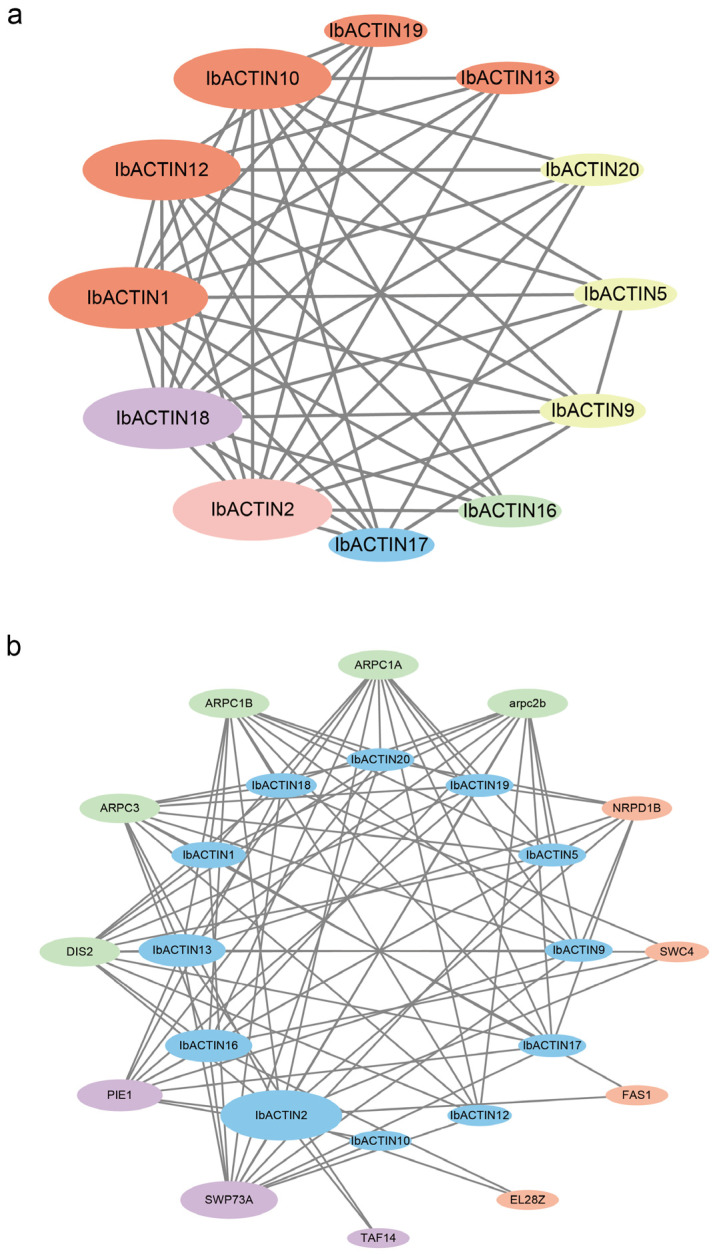
ACTINs are structural proteins widely distributed in plants. They are the main components of microfilaments and participate in many crucial physiological activities, including the maintenance of cell shape and cytoplasmic streaming. Meanwhile, ACTIN, as a housekeeping gene, is widely used in qRT-PCR analyses of plants. However, ACTIN family genes have not been explored in the sweet potato. In this study, we identified 30, 39, and 44 ACTINs in the cultivated hexaploid sweet potato (Ipomoea batatas, 2n = 6x = 90) and its two diploid relatives, Ipomoea trifida (2n = 2x = 30) and Ipomoea triloba (2n = 2x = 30), respectively, via analysis of their genome structure and by phylogenetic characterization. These ACTINs were divided into six subgroups according to their phylogenetic relationships with Arabidopsis thaliana. The physiological properties of the protein, chromosome localization, phylogenetic relationship, gene structure, promoter cis-elements, protein interaction networks, and expression patterns of these 113 ACTINs were systematically investigated. The results suggested that homologous ACTINs are differentiated in the sweet potato and its two diploid relatives, and play various vital roles in plant growth, tuberous root development, hormone crosstalk, and abiotic stress responses. Some stable ACTINs that could be used as internal reference genes were found in the sweet potato and its two diploid relatives, e.g., IbACTIN18, -20, and -16.2; ItfACTIN2.2, -16, and -10; ItbACTIN18 and -19.1. This work provides a comprehensive comparison and furthers our understanding of the ACTIN genes in the sweet potato and its two diploid relatives, thereby supplying a theoretical foundation for their functional study and further facilitating the molecular breeding of sweet potatoes.

Keywords: ACTIN; I. trifida; I. triloba; abiotic stress; hormone treatment; sweet potato; tissue-specific expression; tuberous root development.


Conflict of interest statement

The authors declare no conflict of interest.

Figures

Figure 1
Figure 1
Chromosomal localization and distribution of ACTINs in I. batatas (a), I. trifida (b), and I. triloba (c). The bars represent chromosomes. The chromosome numbers are displayed on the left side, and the gene names are displayed on the right. Each gene location is shown on the line. Detailed chromosomal location information is listed in Table S1.
Figure 2
Figure 2
Phylogenetic analysis of the ACTIN proteins from four plant species (i.e., I. batatas, I. trifida, I. triloba, and Arabidopsis thaliana). In total, 133 ACTINs were divided into six subgroups (Group I to Group VI) according to the evolutionary distance. The crimson stars, blue circles, yellow triangles, and green rectangles represent the 30 IbACTINs in I. batatas, the 39 ItfACTINs in I. trifida, the 44 ItbACTINs in I. triloba, and the 20 AtACTINs in Arabidopsis thaliana, respectively.
Figure 3
Figure 3
Conserved motifs and analysis of the exon-intron structure of the ACTIN family in I. batatas, I. trifida, and I. triloba. (a) The phylogenetic tree shows that the ACTINs are distributed in six subgroups (left), and the eight conserved motifs are shown in different colors. (b) Conserved domain structures of ACTINs. The blue-green box represents the ACTIN domain. The pink box represents the F-box domain. (c) Exon-intron structures of ACTINs. The green boxes, yellow boxes, and black lines represent the UTRs, exons, and introns, respectively.
Figure 4
Figure 4
cis-element analysis of IbACTINs in I. batatas. The cis-elements were divided into five categories. The intensity of the different colors represents the number of cis-elements in the IbACTIN promoters.
Figure 5
Figure 5
Functional interaction networks of IbACTINs in I. batatas according to their orthologs in Arabidopsis thaliana. The network nodes represent proteins, and the lines represent protein-protein associations. (a) The pink node, green node, blue node, yellow node, orange node, and purple node represent the IbACTINs in Group I, Group II, Group III, Group IV, Group V, and Group VI, respectively. The size of each node represents the number of proteins that interact with each other. The lines represent the interaction among ACTIN proteins. (b) The green node, orange node, and purple node represent the cell polarity development proteins, DNA transcription and translation proteins, and regulation of flower development proteins, respectively. The lines represent the interactions of the ACTINs and other proteins.
Figure 6
Figure 6
Gene expression patterns of the IbACTINs of Xushu18 in different tissues of I. batatas (shoot tip, petiole, leaf, stem, fibrous root, pencil root, and storage root).
Figure 7
Figure 7
Gene expression patterns of ItfACTINs (a) and ItbACTINs (b) in the flower bud, flower, leaf, stem, root1, and root2 of I. trifida and I. triloba, as determined by RNA-seq. The log2 (FPKM+1) values are shown in the boxes.
Figure 8
Figure 8
Gene expression patterns of IbACTINs in different developmental stages of the root as determined by RNA-seq. F, fibrous root (diameter of approximately 1 mm); D1, initial storage root (diameter of approximately 1 cm); D3, storage root (diameter of approximately 3 cm); D5, storage root (diameter of approximately 5 cm); D10, storage root (diameter of approximately 10 cm).
Figure 9
Figure 9
Gene expression patterns of ACTINs in response to different phytohormones (ABA, GA, IAA, and BAP) in I. trifida (a) and I. triloba (b) as determined by RNA-seq. The log2 (FPKM+1) values are shown in the boxes.
Figure 10
Figure 10
Gene expression patterns of IbACTINs under drought and salt stresses as determined by RNA-seq. (a) Expression of IbACTINs under PEG treatment in a drought-tolerant variety, i.e., Xu55-2. (b) Expression of IbACTINs under NaCl treatment in a salt-sensitive variety, i.e., Lizixiang, and a salt-tolerant line, i.e., ND98. The log2 (FPKM) values are shown in the boxes.
Figure 11
Figure 11
(ac) Gene expression patterns of ACTINs under abiotic stresses (cold, heat, salt, and drought) in I. trifida, as determined by RNA-seq. (df) Gene expression patterns of ACTINs under abiotic stresses (cold, heat, salt, and drought) in I. triloba, as determined by RNA-seq. COCO, COLD, HECO, HEAT, DSCO, NACL, and MANN represent the cold control at 28/22-deg C day/night experiment, cold stress at 10/4-deg C day/night experiment, heat control at 28/22-deg C day/night experiment, heat stress at 35/35-deg C day/night experiment, drought and salt control, NaCl salt stress experiment, and mannitol drought stress experiment, respectively. The log2 (FPKM+1) values are shown in the boxes.

References

    1. Jürgens G. Cytokinesis in higher plants. Annu. Rev. Plant Biol. 2005;56:281–299. doi: 10.1146/annurev.arplant.55.031903.141636. - DOI - PubMed
    1. Mascarenhas J.P. Molecular mechanisms of pollen tube growth and differentiation. Plant Cell. 1993;5:1303–1314. doi: 10.2307/3869783. - DOI - PMC - PubMed
    1. Baluska F., Hlavacka A., Samaj J., Palme K., Robinson D.G., Matoh T., McCurdy D.W., Menzel D., Volkmann D. F-actin-dependent endocytosis of cell wall pectins in meristematic root cells. Insights from brefeldin A-induced compartments. Plant Physiol. 2002;130:422–431. doi: 10.1104/pp.007526. - DOI - PMC - PubMed
    1. Barrero R.A., Umeda M., Yamamura S., Uchimiya H. Arabidopsis CAP regulates the actin cytoskeleton necessary for plant cell elongation and division. Plant Cell. 2002;14:149–163. doi: 10.1105/tpc.010301. - DOI - PMC - PubMed
    1. Gu Y., Fu Y., Dowd P., Li S., Vernoud V., Gilroy S., Yang Z. A Rho family GTPase controls actin dynamics and tip growth via two counteracting downstream pathways in pollen tubes. J. Cell Biol. 2005;169:127–138. doi: 10.1083/jcb.200409140. - DOI - PMC - PubMed

LinkOut - more resources