. 2023 Jun 30;24(13):10973.
doi: 10.3390/ijms241310973.

Pathophysiology of Cerebellar Degeneration in Mitochondrial Disorders: Insights from the Harlequin Mouse

Affiliations

Pathophysiology of Cerebellar Degeneration in Mitochondrial Disorders: Insights from the Harlequin Mouse

Miguel Fernández de la Torre et al. Int J Mol Sci. .

Abstract

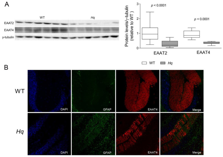
By means of a proteomic approach, we assessed the pathways involved in cerebellar neurodegeneration in a mouse model (Harlequin, Hq) of mitochondrial disorder. A differential proteomic profile study (iTRAQ) was performed in cerebellum homogenates of male Hq and wild-type (WT) mice 8 weeks after the onset of clear symptoms of ataxia in the Hq mice (aged 5.2 ± 0.2 and 5.3 ± 0.1 months for WT and Hq, respectively), followed by a biochemical validation of the most relevant changes. Additional groups of 2-, 3- and 6-month-old WT and Hq mice were analyzed to assess the disease progression on the proteins altered in the proteomic study. The proteomic analysis showed that beyond the expected deregulation of oxidative phosphorylation, the cerebellum of Hq mice showed a marked astroglial activation together with alterations in Ca2+ homeostasis and neurotransmission, with an up- and downregulation of GABAergic and glutamatergic neurotransmission, respectively, and the downregulation of cerebellar "long-term depression", a synaptic plasticity phenomenon that is a major player in the error-driven learning that occurs in the cerebellar cortex. Our study provides novel insights into the mechanisms associated with cerebellar degeneration in the Hq mouse model, including a complex deregulation of neuroinflammation, oxidative phosphorylation and glutamate, GABA and amino acids' metabolism.

Keywords: GABA; Harlequin mouse; OXPHOS disorders; ataxia; complex I; glutamate; long-term depression; mitochondrial diseases.


Conflict of interest statement

The authors declare no conflict of interest. The funders had no role in the design of the study; in the collection, analyses, or interpretation of data; in the writing of the manuscript; or in the decision to publish the results.

Figures

Figure 1
Figure 1
Glutamate transporters. (A) Representative Western blot and densitometry analysis of glutamate transporters EAAT2 and EAAT4 in the cerebellum of WT and Hq mice. γ-tubulin was used as loading control. Data (mean, interquartile range and min and max values) are expressed relative to the WT group. p-Values for differences between WT and Hq groups (Mann–Whitney U test) are shown above the graph. (B) Representative immunofluorescence of GFAP (green), EAAT4 (red) and nuclei staining with DAPI (blue) in cerebellar tissue of WT and Hq mouse. Abbreviations for groups: WT, wild type; Hq, Harlequin.
Figure 2
Figure 2
GABA transporters. Representative Western blot and densitometry analysis of GABA transporters GAT-3 and VGAT in the cerebellum of WT and Hq mice. γ-tubulin was used as loading control. Data (mean, interquartile range and min and max values) are expressed relative to the WT group. p-Values for differences between WT and Hq groups (Mann–Whitney U test) are shown above the graph. Abbreviations for groups: WT, wild type; Hq, Harlequin.
Figure 3
Figure 3
Glutamate receptors and Ca2+ homeostasis-related proteins. Representative Western blot and densitometry analysis of (A) glutamate receptors mGluR1 and GluRδ2 and (B) Ca2+ homeostasis-related proteins, Homer protein homolog 3 (HOMER-3), inositol 1,4,5-triphosphate receptor type 1 (IP3R), myristoylated alanine-rich C-kinase substrate phosphorylated at Serine 152/156 (pMARCKS) and subunit 2 of the glutamate ionotropic receptor AMPA type phosphorylated at Serine 880 (pGRIA2) in the cerebellum of WT and Hq mice. γ-tubulin was used as loading control. Data (mean, interquartile range and min and max values) are expressed relative to the WT group. p-Values for differences between WT and Hq groups (Mann–Whitney U test) are shown above the graph. Abbreviations for groups: WT, wild type; Hq, Harlequin.
Figure 4
Figure 4
Aquaporin 4. Representative Western blot and densitometry analysis of aquaporin 4 in the cerebellum of WT and Hq mice. γ-tubulin was used as loading control. Data (mean, interquartile range and min and max values) are expressed relative to the WT group. Mann–Whitney U test, significantly different compared to WT. Abbreviations for groups: WT, wild type; Hq, Harlequin.

Similar articles

Cited by

References

    1. Gorman G.S., Chinnery P.F., DiMauro S., Hirano M., Koga Y., McFarland R., Suomalainen A., Thorburn D.R., Zeviani M., Turnbull D.M. Mitochondrial diseases. Nat. Rev. Dis. Prim. 2016;2:16080. doi: 10.1038/nrdp.2016.80. - DOI - PubMed
    1. Ng Y.S., Turnbull D.M. Mitochondrial disease: Genetics and management. J. Neurol. 2016;263:179–191. doi: 10.1007/s00415-015-7884-3. - DOI - PMC - PubMed
    1. Fassone E., Rahman S. Complex I deficiency: Clinical features, biochemistry and molecular genetics. J. Med. Genet. 2012;49:578–590. doi: 10.1136/jmedgenet-2012-101159. - DOI - PubMed
    1. Gorman G.S., Schaefer A.M., Ng Y., Gomez N., Blakely E.L., Alston C.L., Feeney C., Horvath R., Yu-Wai-Man P., Chinnery P.F., et al. Prevalence of nuclear and mitochondrial DNA mutations related to adult mitochondrial disease. Ann. Neurol. 2015;77:753–759. doi: 10.1002/ana.24362. - DOI - PMC - PubMed
    1. Fiedorczuk K., Sazanov L.A. Mammalian Mitochondrial Complex I Structure and Disease-Causing Mutations. Trends Cell Biol. 2018;28:835–867. doi: 10.1016/j.tcb.2018.06.006. - DOI - PubMed